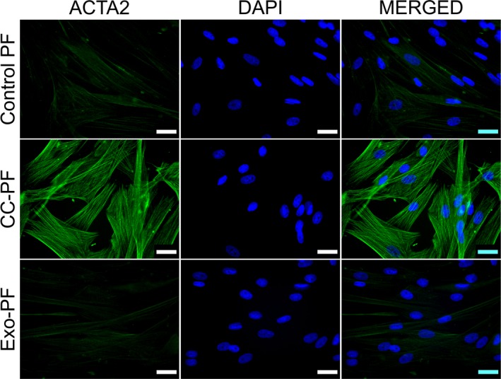
Fig. 1.

Immunostaining in canine PFs for ACTA2 protein. Images show well‐defined and organised long intracytoplasmic ACTA2 filaments in CC‐PF 96 h after coculture compared with control PF and Exo‐PF groups. IF representative images using at least two biological replicates were taken under identical microscope and camera settings. Scale bars represent 25 μm
