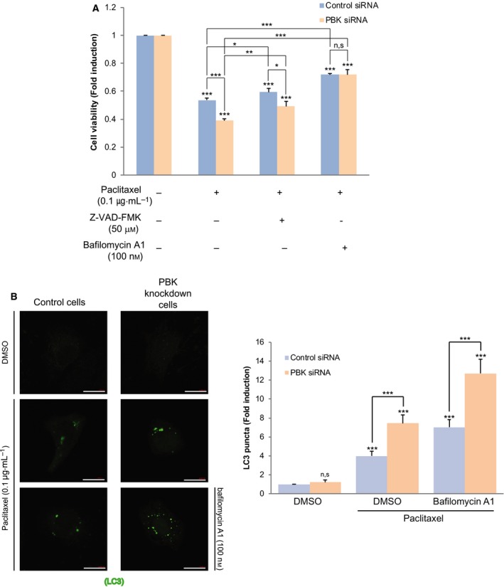
Fig. 2.

Depletion of PBK increases autophagic cell death in response to paclitaxel. (A) Control siRNA cells or PBK siRNA cells H460 cells were treated with DMSO, and paclitaxel (0.1 μg·mL−1) alone or in combination with Z‐VAD‐FMK (50 μm) or bafilomycin A1 (100 nm) for 24 h, and then, cell viability was examined. (B) Control cells or PBK knockdown cells were transfected with GFP‐LC3 construct. Twenty‐four hours after transfection, cells were incubated with paclitaxel (0.1 μg·mL−1) alone or paclitaxel plus bafilomycin A1 (100 nm) for 24 h. GFP‐LC puncta (green) was observed by confocal microscopy. Scale bar, 20 μm. Representative images of 63× magnification from three independent experiments are indicated. *P < 0.05; **P < 0.01; and ***P < 0.001 compared to controls. N.S, nonsignificant. Statistical analysis was performed by one‐way ANOVA.
