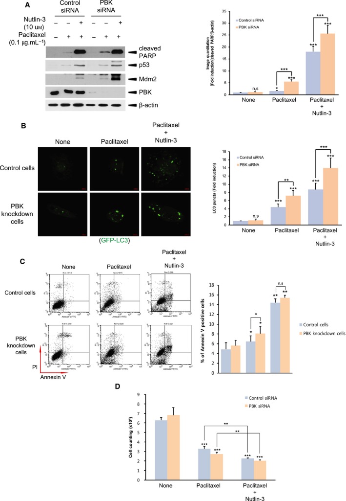
Fig. 6.

Inhibition of interaction of Mdm2 with p53 promotes both autophagy and apoptosis during paclitaxel treatment. (A) Control siRNA cells or PBK siRNA cells were treated with DMSO, and paclitaxel (0.1 μg·mL−1) alone or in combination with nutlin‐3 (10 μm) for 24 h. Cell lysate was subjected immunoblotting using indicated antibody. (B) Control cells or PBK knockdown cells were transfected with GFP‐LC and 24 h after transfection, cells were incubated DMSO, and paclitaxel (0.1 μg·mL−1) alone or in combination with nutlin‐3 (10 μm) for 24 h. GFP‐LC puncta was observed by confocal microscopy. Representatives of 63× magnification from three independent experiments are indicated. Scale bar, 20 μm. (C) Control cells or PBK knockdown cells were treated with DMSO, and paclitaxel (0.1 μg·mL−1) alone or in combination with nutlin‐3 (10 μm) for 24 h. Apoptosis was analyzed by Annexin V‐FITC and propidium iodide staining, and subsequent flow cytometry. Representatives of three independent experiments and graph for quantitation of Annexin V‐positive cells are shown. (D) Control siRNA cells or PBK siRNA cells were incubated with DMSO, and paclitaxel (0.1 μg·mL−1) alone or in combination with nutlin‐3 (10 μm) for 24 h. Cell counting was done by colony‐forming assay. *P < 0.05; **P < 0.01; and ***P < 0.001 compared to controls. Results are indicated as the mean ± SD for at least three independent experiments in duplicates. Statistical analysis was done by two‐tailed Student's t‐test.
