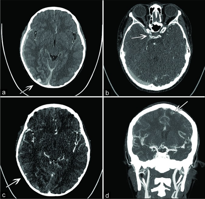
Figure 1:

(a) The right-sided subdural hematoma with large mycotic aneurysm at the periphery of the right occipital lobe, (b) the right cavernous ICA aneurysm, (c) the right parietal temporal junction aneurysm (d) Small aneurysm of the left frontal lobe.
