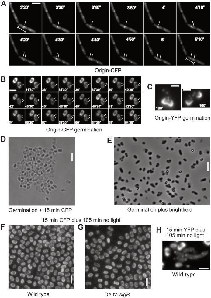
Fig. 1.
Blue light excitation arrests cell growth in Bacillus subtilis. a) Exponentially growing B. subtilis cells carrying a lacO/LacI-CFP fluorescent repressor-operator (FROS) tag (strain PG26) were imaged with 445 nm laser excitation at 10 s intervals. Cell growth ceased after 3 min, but one origin separation event can be observed (white dashes). b-e) B. subtilis spores were heat-induced and germinated in germination medium. Shown are cells imaged at the indicated times after germination. In b), cells of strain PG26 were subjected to 15 exposures (0.5 s each) with CFP excitation, every minute, 45 min after the induction of germination, which prevented germination in the majority of spores. Cells were imaged 105 min after the induction of germination shown are time intervals between minute 31 and 65 of a representative experiment. Cells in C) carrying a TetR-YFP/tetO FROS system near the origin region (KS188) were subjected to 15 exposures (0.5 s each) with YFP excitation, every minute, 45 min after the induction of germination, allowing germination and subsequent cell growth of a majority of spores. Cells were imaged 120 min after the induction of germination. Spore coats in b and C show up by intensive fluorescence. d-e) Spores devoid of a FROS system were subjected to 15 exposures with CFP excitation (0.5 s each) for the first 15 min (d) or to a similar treatment using bright field illumination (e), followed by further incubation for 105 min without light. Phase-bright cells are non-germinated spores, dark cells have germinated. f) Spores of strain PG26, or g) ΔsigB mutant spores, carrying the LacI-CFP/lacO FROS system were subjected to 15 exposures with CFP excitation (0.5 s each) for the first 15 min, followed by further incubation for 105 min without light (analogous to d-e). h) Spores of strain KS188 (TetR-YFP/tetO system) were subjected to 15 exposures with YFP excitation (0.5 s each) for the first 15 min, followed by further incubation for 105 min without light (analogous to d-g) to continue germination. Note that 75% of all cells (n = 250) had already divided at this time point. White bars 2 μm

