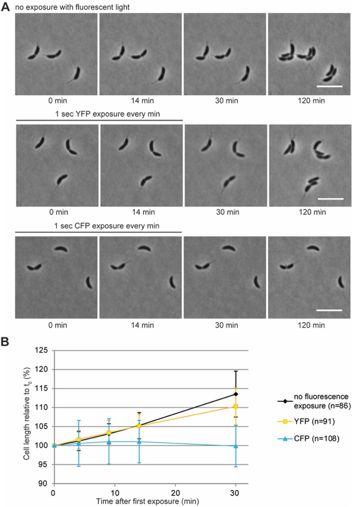
Fig. 5.
CFP exposure has a bacteriostatic effect on C. crescentus cells. (a) Phase contrast images of C. crescentus cells imaged on 1% PYE agarose pads show that cells that were exposed to white light only (top row) or additionally to YFP excitation (1 s per burst, 16 burst in total with a time interval of 1 min) (middle row) grew both during the initial phase of imaging (at 1 min intervals) and during the recovery period, in which they were imaged by phase contrast microscopy only every 15 min. As a result, cell division occurred in all cells during the first 120 min of the experiment. In contrast, C. crescentus cells that were exposed to 16 pulses of CFP excitation (during the first 15 min of the experiment) (bottom row) halted growth and did not produce daughter cells. White bars 5 µm. (b) Quantification of the growth of individual cells during exposure to white light only or with additional YFP or CFP exposures shows that CFP exposure inhibits cell growth already after a few (less than 5) bursts, whereas cells that are exposed to YFP or white light do not show any growth defect. No recovery of growth was seen within the first 15 min of recovery. Data are the mean of 3 independent experiments. Error bars represent the standard deviation

