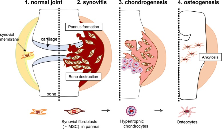
Fig. 6.
Schematic representation of the stages of RA in D1BC mouse model [16]. 1: Synovial membranes, which consist of one to three cell layers, are the origin of synovial fibroblasts. 2: Joint inflammation induces aggressive proliferation of synovial fibroblasts, followed by bone erosion and destruction in the inflamed joint. Synovial fibroblasts share cell lineage markers of MSCs with pre-hypertrophic chondrocytes in bColII-D1BC mice. Because the most definitive feature of synovial fibroblasts in pannus is the expression of Col10a1 mRNA, but not its transcribed protein, ColX, these are defined as pre-hypertrophic chondrocytes. Thus, a subpopulation of synovial fibroblasts (B7.1, Col2a1, Runx2, Sox9, Col10a1, and Osx) differentiates into ColX-negative hypertrophic chondrocytes. 3: Synovial fibroblasts differentiate into hypertrophic chondrocytes by hypertrophic cartilage remodeling in the process of endochondral bone formation. 4: When hypertrophic chondrocytes terminally differentiate into osteochondrocytes, the expression of ColI is observed instead of ColX. A failure of cartilage remodeling leads to aberrant bone formation by ossified chondrocytes, resulting in bony ankylosis

