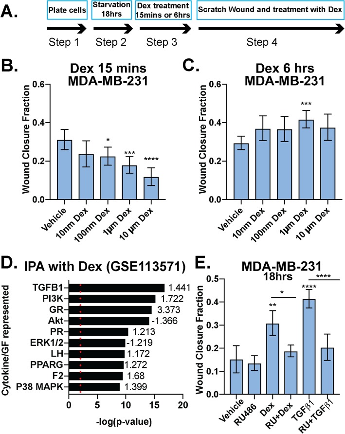
Fig. 1.
Dexamethasone either inhibits or promotes breast cancer cell migration in a time-dependent manner. a Schematic of protocol used for b and c. MDA-MB-231 cells were pretreated with increasing doses of Dex for either 15 min (b) or 6 h (c) and Dex-induced cell migration was measured by the degree of scratch wound closure at 18 h. The mean of three field images from each of the three biological replicates experiments is shown ± standard deviation (SD). Fraction of wound area closure of MDA-MB-231 cells was determined ImageJ. Statistical significance was assessed by one-way ANOVA and Dunnett’s post hoc for comparison within groups vs. vehicle treatment. (*, p < 0.05; **, p < 0.01; ***, p < 0.001; ****, p < 0.0001). d Ingenuity Pathway Analysis (IPA) for MDA-MB-231 cells treated with 100 nM Dex from GSE113571 [12]. p value plot shows GR-mediated activation of cancer-relevant signaling pathways and their respective z-scores of activation/inhibition, including the TGFβ1 and p38 MAPK pathways. The top 10 most significant pathways for cytokine/growth factor (GF) signaling are shown. Red dotted line indicates the significance value of 1.3 (p < .05). e Fraction of wound area closure of MDA-MB-231 cells treated (18 h) with vehicle control, TGFβ1 (10 ng/mL), Dex (1 μM), TGFβ1+Dex, RU486 (1 μM), RU486+TGFβ1, or RU486+Dex. The mean of three fields from each of the three biological replicate experiments is shown ± SD. Statistical significance was assessed by one-way ANOVA and Tukey post hoc for comparison within groups. (*, p < 0.05, **, p < 0.01, ****, p < 0.0001)

