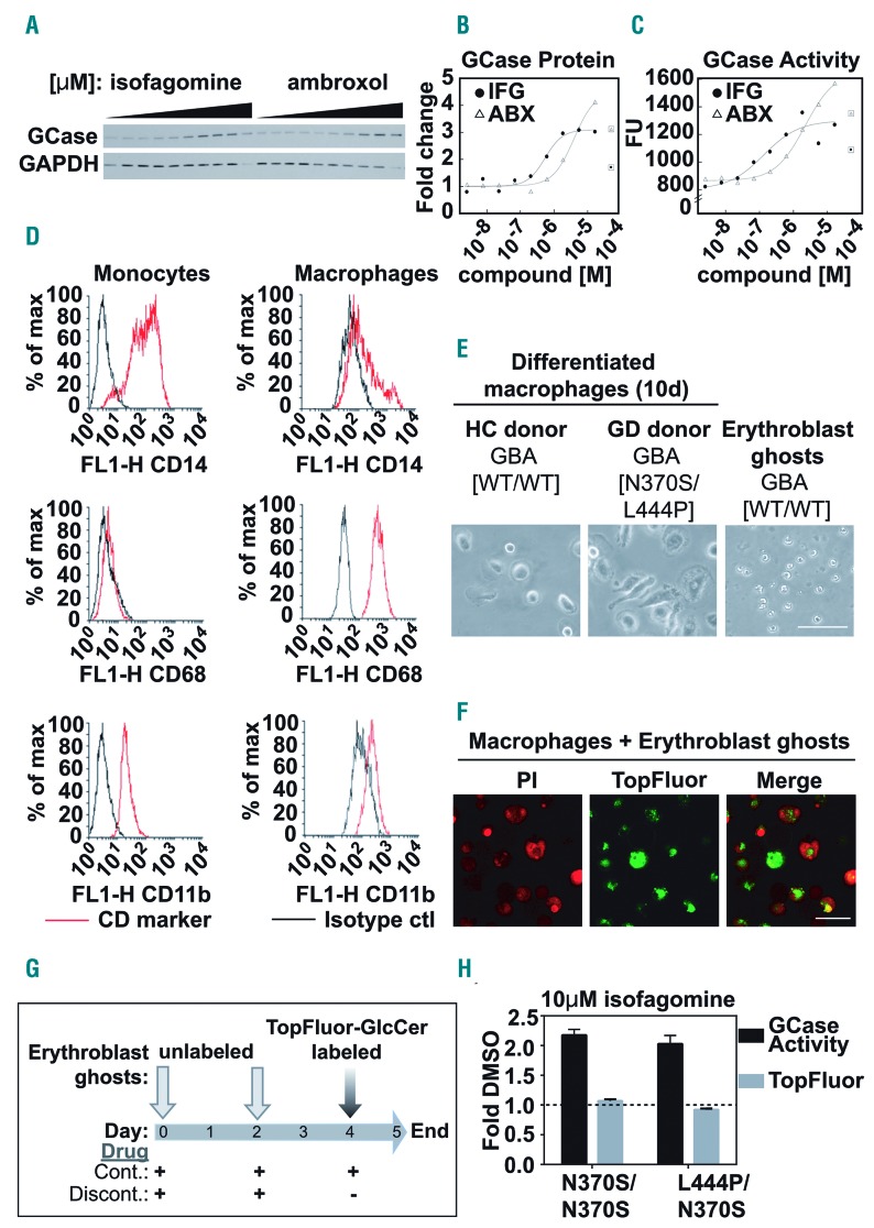
Figure 1.
Patient blood monocytic cell-derived macrophage model to assess the functional impact of glucocerebrosidase-specific inhibitory chaperone compounds. (A-C) Fibroblasts from a Gaucher disease (GD) patient [GBA1 N370S/del] were treated with increasing doses (0-50 μM) of the glucocerebrosidase (GCase) inhibitors isofagomine (IFG) or ambroxol (ABX) for 6 days before the cells were harvested. (A) Western blot of the protein levels of GCase and GAPDH, as a loading control, in whole cell lysates. (B) Dose-response curves of densitometrically quantified GCase protein levels. Boxed points are outliers removed due to observable toxicity. (C) Dose-response curves of GCase activity using whole cell lysates from ambroxol- or isofagomine-treated GD fibroblasts. Boxed points are outliers removed due to observable toxicity. (D-G) Patient-derived monocytes were isolated using a Percoll gradient and CD14+ magnetic beads and were then differentiated into patient blood monocytic cell (PBMC)-derived macrophages using granulocyte-macrophage colony-stimulating factor. Erythroblast ghosts were generated by hypo-osmotic lysis. Unlabeled erythroblast ghosts were added to the macrophages for phagocytosis at assay set up (day 0) and at 48 h (day 2), to saturate the intracellular glycolipid pool. Twenty-four hours before the assay readout (day 4) erythroblast ghosts labeled with TopFluor-glucosylceramide (GlcCer) were added to the macrophages. Remaining TopFluor-GlcCer levels in PBMC-derived macrophages were read out at 485/528 nm using a spectrophotometer.12 (D) Fluorescence activated cell sorting analysis showing enrichment of the CD68+ population of differentiated macrophages compared with CD14/CD11b+ monocytic precursors. (E) Transmitted light micrographs showing representative PBMC-derived macrophages from a healthy control (HC) donor (left) and a GD patient (middle) and erythroblast ghosts (right). (F) Confocal micrographs of propidium iodide (PI)-labeled fixed PBMC-derived macrophages (red) and TopFluor-labeled erythroblast ghosts (green) after incubation for 24 h with TopFluor-GlcCer-labeled erythroblast ghosts. (G) Schematic representation of erythroblast ghost delivery and compound treatment protocols. On day 4, compounds were either (i) replenished as part of a continuous protocol (Cont.), or (ii) removed for the 24 h period of TopFluor-GlcCer-labeled erythroblast ghost delivery in a discontinuous protocol (Discont.). (H) Two different GD PBMC-derived macrophage samples were exposed to 10 μM isofagomine in the Continuous protocol and GCase activity (black bars) and TopFluor-GlcCer (gray bars) were measured and expressed as fold change compared to those of the samples exposed to dimethylsulfoxide (DMSO), the vehicle control (dotted line at 1). Scale bar in (E) and (F) = 50 μM.

