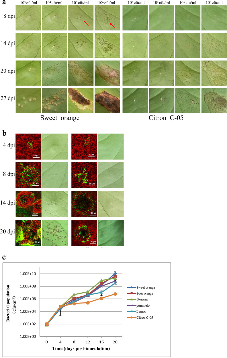
Fig. 3. Infection phenotypes of sweet orange and Citron C-05 following infiltration inoculation.
Xcc bacterial suspensions at different concentrations (103, 104, 105, and 106 cfu/ml) were infiltrated from the abaxial surface into fully expanded young leaves of the six citrus biotypes, and disease symptoms were monitored and documented from 1 to 27 dpi. Images of sweet orange and Citron C-05 are shown here. Images of the other biotypes are shown in Supplementary Fig. S2. a Xcc infection symptoms of sweet orange and Citron C-05 leaves infiltrated with the four indicated Xcc concentrations at the four indicated time points. b Visualization of Xcc after infiltration (at 104 cfu/ml) of the leaves of sweet orange and Citron C-05 by CLSM at the indicated time points. The infection symptoms of the same leaves were photographed and are shown in the right panels. c Quantification of Xcc in the leaves of the six citrus biotypes infiltrated with 104 cfu/ml Xcc

