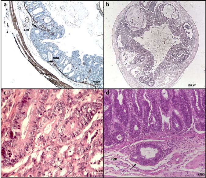
Figure 1.
Histological sections of ileocecal regions of Dexamethasone-treated SCID mice infected with different C. parvum isolates. (a) C. parvum IOWA after 107 days post-infection (PI): presence of an invasive adenocarcinoma reaching the submucosa (sm) with an interruption (arrows) of the muscularis mucosae (mm) (immunohistochemical stain for alpha smooth muscle actin). Bar, 70 μm. (b) C. parvum DID after 62 days PI: presence of an adenocarcinoma invading the submucosa (hematoxylin and eosin staining). Bar, 250 μm. (c) C. parvum TUM1, after 19 days PI: high grade intraepithelial neoplasia characterized by epithelial atypia and associated with the presence of numerous parasites inside the glands (arrow) (hematoxylin and eosin staining). Bar, 12 μm. D) C. parvum CHR after 15 days PI: development of an adenocarcinoma (arrow) in the submucosa (sm) (hematoxylin and eosin staining). Bar, 30 μm.

