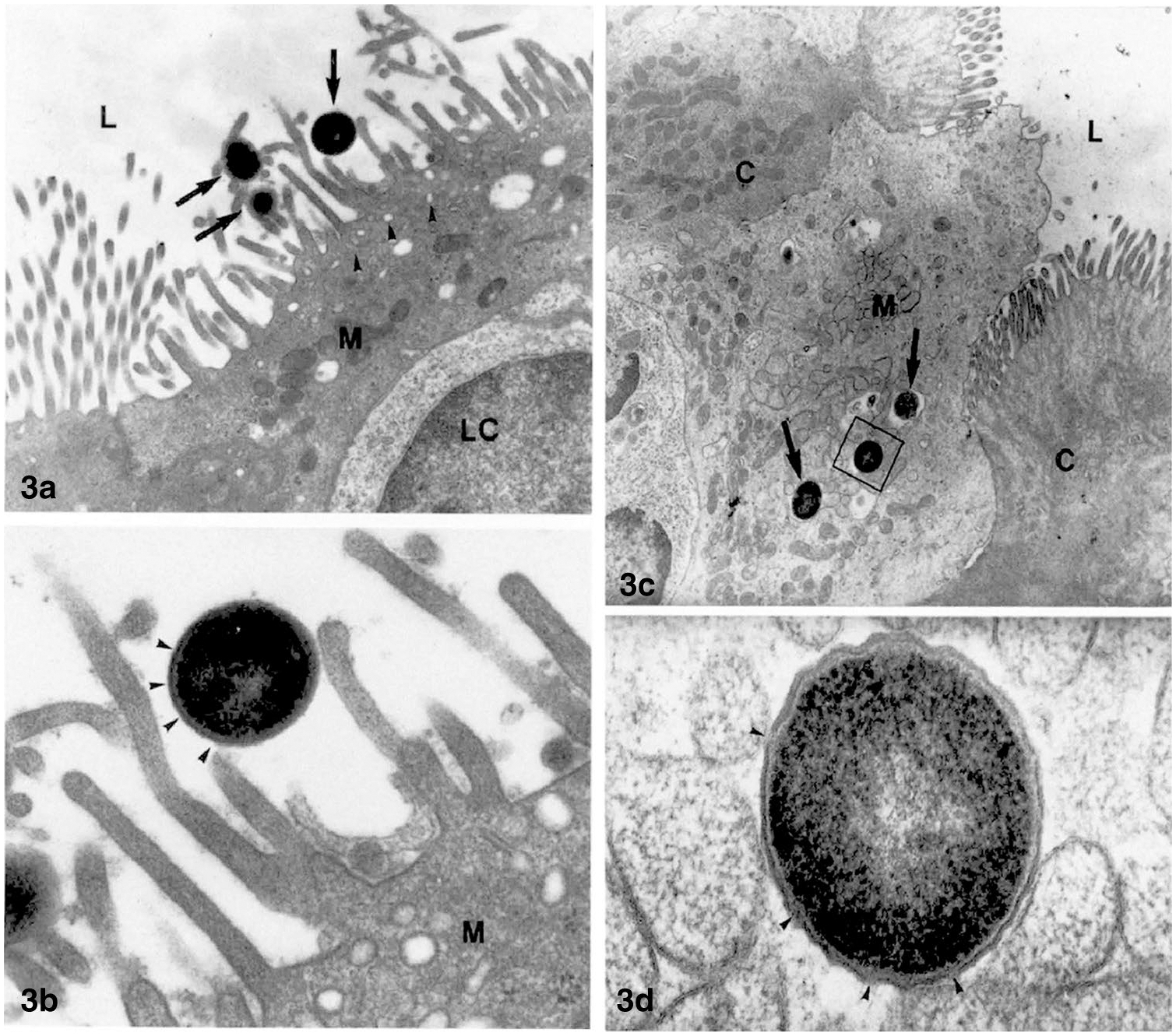
Fig. 3.

Transmission electron microscopic images of the entry of intact bacteria into M-cells overlying the Peyer’s patches in rabbit ileum. Isolated loops of ileum were incubated with a suspension of S. pneumoniæ R36a prior to harvesting the tissue. Caption from 61 “a TEM image of a M-cell (M) from 30 min treated PP. The cell shows the typical morphology: short microvilli, many pinocytotic vesicles (arrowheads) in the apical cytoplasm and a lymphoid cell (LC) close to the gut lumen (L). Among the microvilli three S. pneumoniae (arrows) are present. × 1000. b Detail of a. The bacterial wall (arrowheads) appears intact without damaged areas. M = M-cell. × 36,360. c Apical portion of a M-cell (M) with some streptococci inside endosomes (arrows), from a 60 min treated PP. The M-cell protrudes into the intestinal lumen (L) and joins the adjacent columnar cells (C). × 7870. d Enlargement of the square in c. The pneumococcus shows some broken areas in its wall (arrowheads). × 77,780.” Reproduced from [61] with permission
