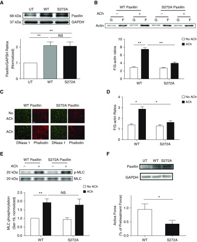
Figure 4.
Paxillin phosphorylation at Ser-272 modulates actin polymerization, but not myosin light chain phosphorylation, at Ser-19 in smooth muscle. (A) Extracts of untransfected (UT) cells and cells expressing wild-type (WT) or S272A paxillin were evaluated by immunoblot analysis. Data are mean values of experiments from four batches of cell culture from three donors. Error bars represent SD. (B) HASM cells expressing WT or S272A paxillin were stimulated with ACh (10−4 M, 5 min), or left unstimulated. F-actin/G-actin ratios were evaluated using the fractionation assay. Data are mean values of four batches of cell culture from three donors. Error bars indicate SD. (C and D) Fluorescent images illustrating the effects of S272A paxillin on F/G-actin ratios in HASM cells. Expression of S272A paxillin attenuates the ACh (10−4 M, 5 min) -induced F/G-actin ratios. Data are means ± SD (n = 24–26 images from three independent experiments). Scale bar: 30 μm. (E) Smooth muscle cells expressing recombinant paxillin were stimulated with ACh (10−4 M, 5 min) or left unstimulated. MLC phosphorylation at Ser-19 was determined by IB. Data are mean values of four batches of cell culture from three donors. Error bars represent SD. (F) Immunoblots showing the expression of WT or S272A paxillin in human bronchial tissues. Blots are representative of three identical experiments. Contractile response of human bronchial rings was evaluated, followed by reversible permeabilization to introduce constructs for WT or S272A paxillin. Active force (ACh, 10−4 M, 5 min) was compared before and after the introduction. Data are mean values of six samples from three donors. Error bars indicate SD. *P < 0.05 and **P < 0.01. One-way ANOVA was used for statistical analysis of A. Two-way ANOVA was used for statistical analysis of B, D, and E. Student’s t test was used for statistical analysis of F.

