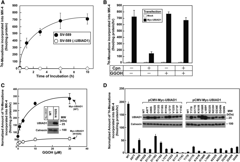
Fig. 2.
SCD-associated variants of UBIAD1 are defective in MK-4 synthetic activity. A: SV-589 and SV-589 (ΔUBIAD1) cells were set up on day 0 at 2.5 ± 105 cells per 60 mm dish in medium A containing 5% FCS. On day 1, the cells were switched to medium A supplemented with 5% FCS and 10 μM compactin. Following incubation for 16 h at 37°C, cells were refed identical medium containing 50 nM [3H]menadione (2.5 μCi per reaction) and 100 μM GGOH for the indicated period of time. Following incubations, the cells were washed, lysed in 0.1 N NaOH, and lipids were extracted as described in the Experimental Procedures. The amount of [3H]menadione incorporated into MK-4 was determined by TLC, followed by scintillation counting. The values represent the mean of triplicate reactions (standard error). B: SV-589 (ΔUBIAD1) cells were set up on day 0 at 4 × 10⁵ cells per 60 mm dish in medium A containing 5% FCS. On day 1, the cells were transfected with 1.5 μg per dish pCMV-Myc-UBIAD1 or empty pcDNA 3.1 vector (Mock) in medium A supplemented with 5% FCS. On day 2, cells were switched to medium A containing 5% FCS in the absence or presence of 10 μM compactin. After incubation for 16 h at 37°C, cells were treated for 5 h with 50 nM [3H]menadione (2.5 μCi per reaction) in the absence or presence of 10 μM compactin and/or 30 μM GGOH as indicated. Cells were subsequently harvested and the incorporation of [3H]menadione into MK-4 was determined as described in A. Values represent the mean of triplicate reactions (standard error). C: SV-589 (ΔUBIAD1) cells were set up on day 0 at 2.5 × 10⁵ cells per 60 mm dish in medium A containing 5% FCS. On day 1, the cells were transfected with 1.5 μg per dish pCMV-Myc-UBIAD1 (WT) or pCMV-Myc-UBIAD1 (N102S) in medium A containing 5% FCS. On day 2, cells were switched to medium A supplemented with 5% FCS and 10 μM compactin. Following incubation for 16 h at 37°C, the cells were refed identical medium containing 50 nM [3H]menadione (2.5 μCi per reaction) and the indicated concentrations of GGOH. Following incubation for 5 h, cells were harvested and incorporation of [3H]menadione into MK-4 was determined as described in A. Aliquots of membranes (30 μg protein per lane) isolated from transfected cells were subjected to SDS-PAGE, and immunoblot analysis was carried out with IgG-9E10 (against Myc-UBIAD1) and anti-calnexin IgG (see inset). The band corresponding to Myc-UBIAD1 was quantified using ImageJ software and used to normalize values obtained for [3H]MK-4 synthetic activity. Values represent the mean of triplicate reactions (standard error). D: SV-589 (ΔUBIAD1) cells were set up on day 0 as described in A and transfected on day 2 with 1.5 μg per dish pCMV-Myc-UBIAD1 (WT) or the indicated SCD-associated variant of Myc-UBIAD1 in medium A supplemented with 5% FCS and 10 μM compactin. On day 3, cells received identical medium containing 50 nM [3H]menadione (2.5 μCi per reaction) and 30 μM GGOH. Following incubation for 6 h at 37°C, cells were harvested and incorporation of radioactivity into MK-4 was determined as described in A. Aliquots of membranes (30 μg protein per lane) isolated from transfected cells were subjected to SDS-PAGE, and immunoblot analysis was carried out with IgG-9E10 (against Myc-UBIAD1) and anti-calnexin IgG (see inset). The normalized amount of [3H]MK-4 synthetic activity was determined as described in C. Values represent the mean of triplicate incubations (standard error).

