Fig. 3.
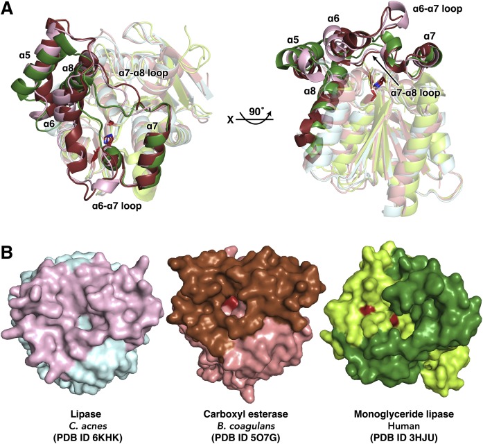
Structural comparison of CAlipase with related proteins. A: The superposition of CAlipase with carboxyl esterase from B. coagulans (core and lid domains are colored in salmon and brown) and monoglyceride lipase from human (core and lid domains are colored in lime and green). The core domain is colored in light color. The conserved catalytic triads (serine, aspartate, and histidine) are indicated as sticks. B: Surface structures of superposed structures in A. The catalytic triad is colored in red and detected under the lid region, but is completely hidden in CAlipase.

