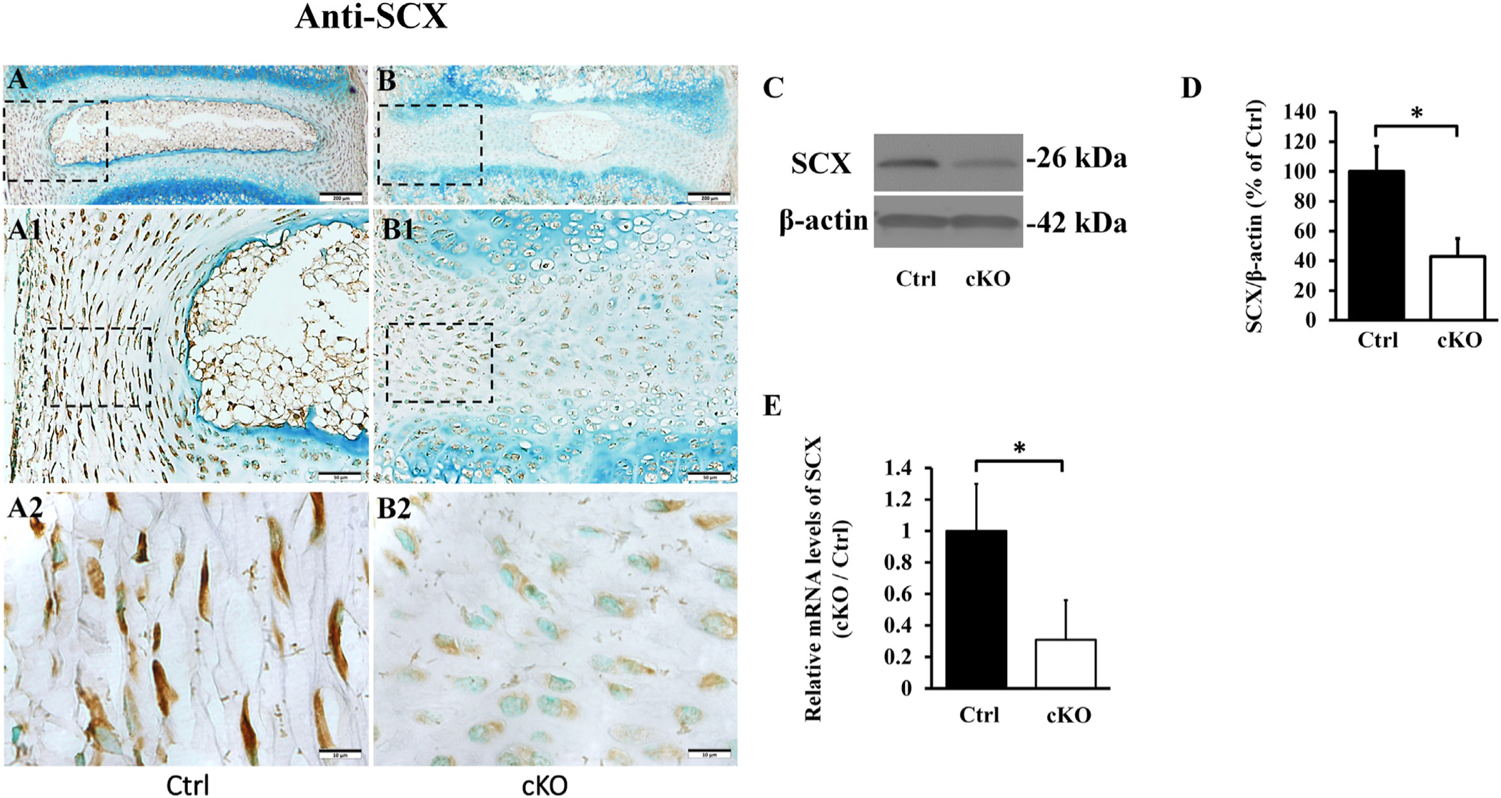
Fig. 10.

Inactivation of Fam20B reduced the expression of SCX in AF. (A, B) IHC detection of SCX in the IVD of 3-week-old control and cKO mice (representative images of at least 3 mice for each group). A1 and B1 were the higher magnification views of the black boxes in A and B, respectively. A2 and B2 were the higher magnification views of the black boxes in A1 and B1, respectively (Bars in the A, B = 200 μm, Bars in the A1, B1 = 50 μm, Bars in the A2, B2 = 10 μm). Note the reduced number of SCX-positive cells in the AF of cKO mice. (C) Anti-SCX Western immunoblotting. (D) Relative levels of SCX protein illustrated in Western immunoblotting analyses (n = 3, * = p < 0.05). The protein level of SCX was markedly lower in AF of cKO mice than in the control. The total proteins were extracted from the AF of 3-week-old mice. (E) SCX mRNA levels detected by QRT-PCR analyses of total mRNAs extracted from the AF of 3-week-old mice; note the reduced level of SCX mRNA level in the AF of cKO mice (n = 6, * = p < 0.05).
