Fig. 2.

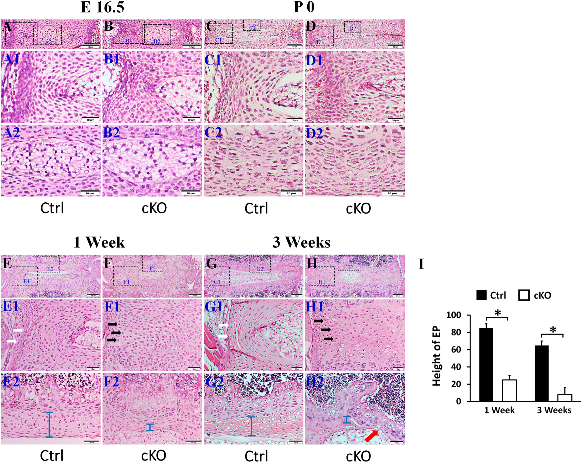
Inactivation of FAM20B caused abnormalities in IVD. H&E staining of mid-coronal (A, B, C, D, E, F, G, H) sections of L3 lumber IVDs of control mice and cKO mice (representative images of at least 3 mice for each group). A1, B1, C1, D1, E1,F1, G1 and H1 were the higher magnification views of larger (left) boxes in A, B, C, D, E,F, G, and H, respectively; A2, B2, C2, D2, E2,F2, G2, and H2 were the higher magnification views of smaller (right) boxes in panels A, B, C, D, E, F, G and H, respectively (Bars in the A, B, C, D, E1, F1, G1, H1, E2, F2, G2, H2 = 50 μm, Bars in the E, F, G, H = 200 μm). (I) Average EP heights in the IVDs of 1-week-old and 3-week-old mice (n = 3). The cKO mice at E 16.5 days and at birth showed mildly malformed IVD, with the AF cells became bigger and rounder. In the 1-and 3-week-old cKO mice, the size of the NP was remarkably smaller and the EP (blue bar) was apparently thinner than the age-matched control mice; in some areas the EP was completely lost (red arrow in H2). The AF cells in 1-and 3-week-old cKO mice lost their normal morphology of spindle shaped cells (white arrows) and were replaced by the rounder chondrocyte-like cells (black arrows).
