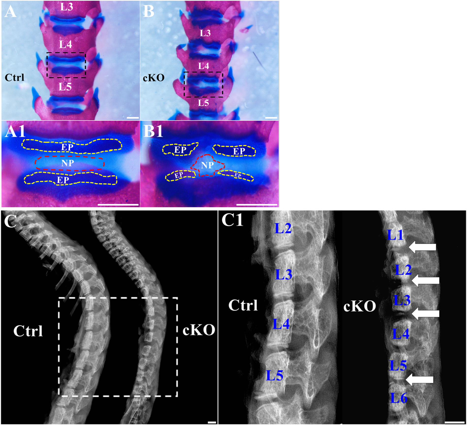
Fig. 3.

Inactivation of FAM20B led to the shrinkage of NP and malformation of EP. (A, B) Alcian blue and alizarin red staining of the spines from 3-week-old control and cKO mice; the representative images (of at least 3 mice for each group) were the anterior views of the L3–L5 region of the spines. A1 and B1 were the higher magnification views of the black boxes in A and B, respectively. The size of NP (the area surrounded by red dashed lines) was reduced in cKO mice and the integrity of EP (the area surrounded by yellow dashed lines) was lost in cKO mice (Bars in the A, B, A1, B1 = 1 mm). (C) Plain X-ray images of the spines from 3-week-old control and cKO mice. C1 was the higher magnification view of white box in panel C. The white arrows indicated the malformed EP in cKO mice (Bars in the C, C1 = 1 mm).
