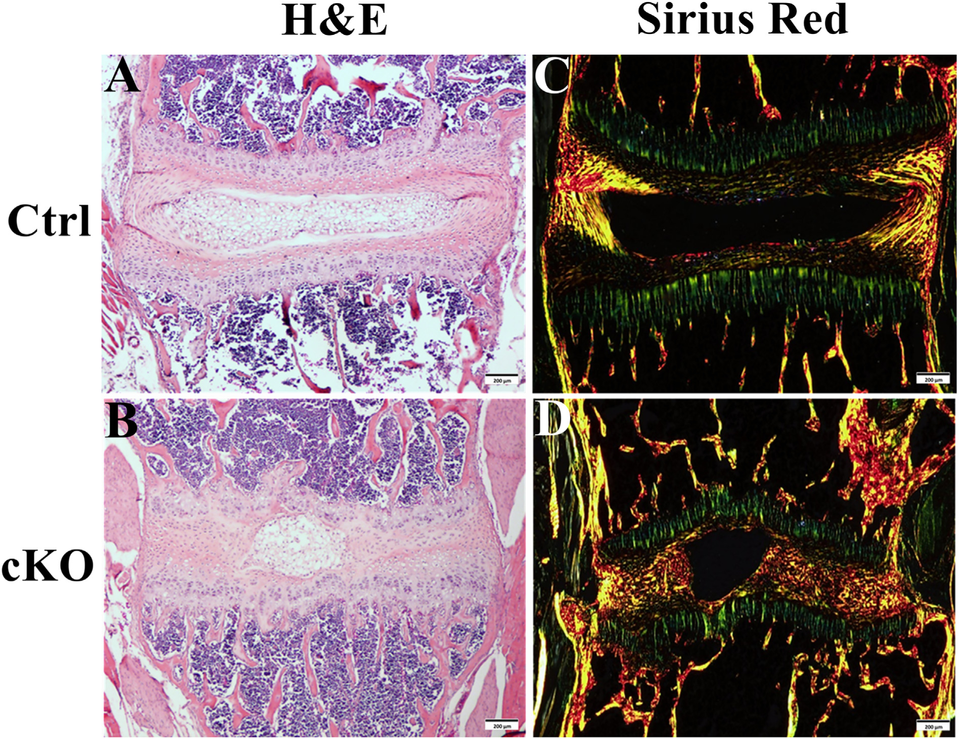
Fig. 5.

Inactivation of FAM20B caused collagen structure changes in AF. (A, B) H&E staining of IVD from 3-week-old control and cKO mice (Bars in the A, B = 200 μm). (C, D) Sirius red staining of IVD in 3-week-old control and cKO mice (Bars in the C, D = 200 μm). A–D was the representative images of at least 3 mice for each group. Note the severe disruption of the collagen structure and thinner and more irregular fibers in the AF of cKO mice. Also note the loss of lamellae in AF of cKO mice.
