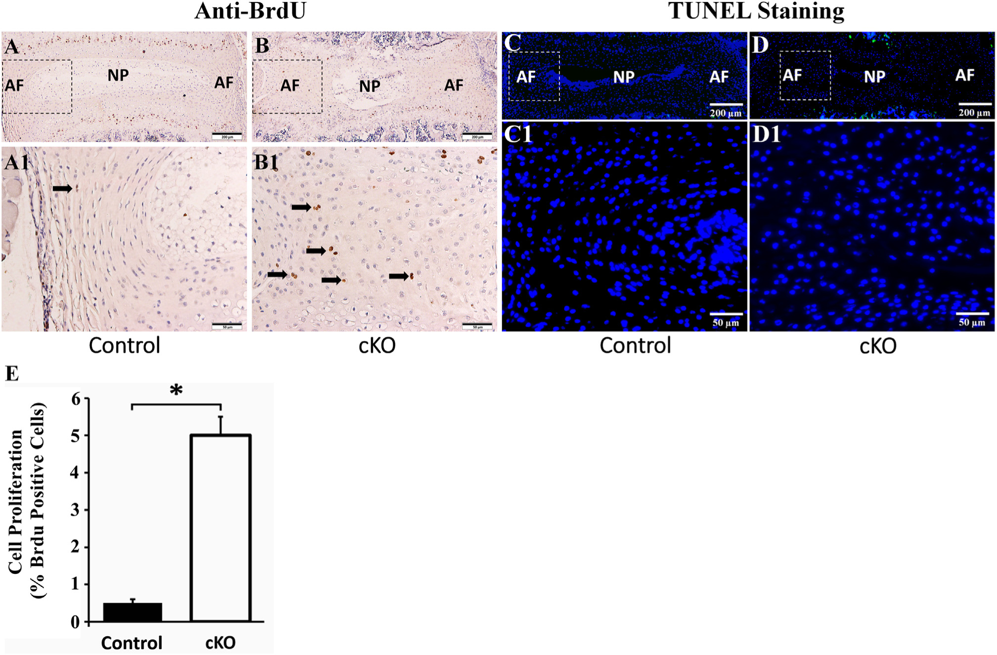
Fig. 7.

Inactivation of FAM20B increased the proliferation of AF cells. (A, B) Anti-BrdU staining of IVDs in 3-week-old control and cKO mice (representative images of 6 mice from each group). A1 and B1 were the higher magnification views of the black boxes in A and B, respectively. The number of BrdU-positive AF cells (indicated by black arrows) in cKO mice was greater than in the control mice (Bars in the A, B = 200 μm, Bars in the A1, B1 = 50 μm). (C, D) TUNEL staining of IVDs in 3-week-old control and cKO mice (representative images of 3 mice from each group). C1 and D1 were the higher magnification views of the white boxes in C and D, respectively. No apoptosis cells were found in the AF of control and cKO mice while positive signals of TUNEL (green) were observed in the growth plates of these mice (Bars in the C, D = 200 μm, Bars in the C1, D1 = 50 μm). (E) Quantitative comparison of cell proliferation rates between AF cells of cKO mice versus the control mice as measured by the BrdU-positive cells. (n = 6; * = p < 0.05).
