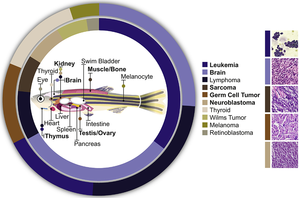
Figure 1. Key Figure. Frequency of Pediatric Cancers and Corresponding Anatomical Location in Zebrafish.

The frequency of childhood (inner circle) and adolescent (outer circle) cancers (as reported in [11,36]) with corresponding anatomical tumor location in zebrafish is illustrated. Zebrafish models have been developed for pediatric leukemia, brain tumors, sarcomas, germ-cell tumors and neuroblastoma (bolded). Representative histology of these zebrafish models is shown on the right. Histology images from: [26] (T-ALL), [39] (CNS-PNET), [62] (ERMS), [71] (Germ-Cell Tumor), and [50] (NB).
