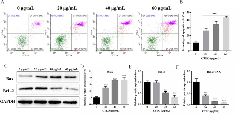
Fig 6. CTEO promoted A549 cells apoptosis in a dose-dependent manner.

A, B. The apoptosis of A549 cells following 48 h of different concentrations CTEO treatment was detected by flow cytometry, and quantification. C-F. The expression of Bax and Bcl-2 protein in A549 cells following 48 h of different concentrations CTEO treatment were detected by western blot analysis, and quantification. Values represent mean ± SD of three independent experiments. *P < 0.05, **P < 0.01, and ***P < 0.001.
