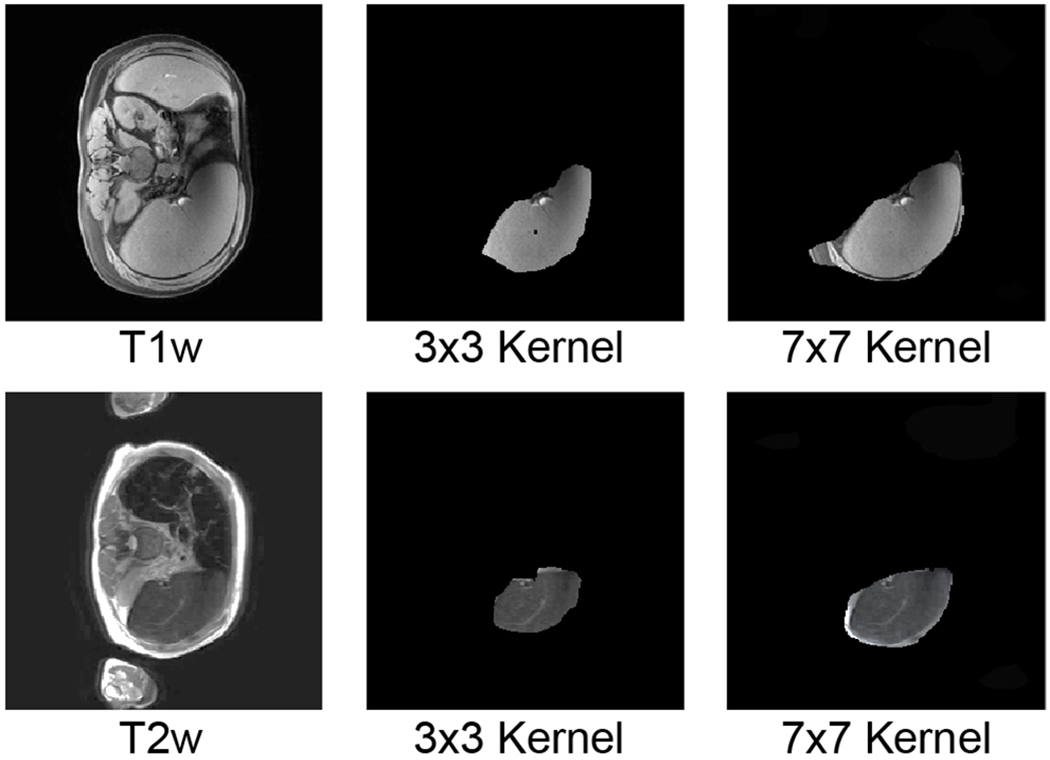
Fig. 2.

This figure compares using large convolutional kernel and small convolutional kernel in splenomegaly segmentation. The upper row indicated a T1w image, while the lower row posed a T2w image. The first column was the original intensity image while the right two columns indicated masked valid field of view (FOV) in skip-connector layers. The larger kernel (7 × 7) has larger valid FOV compared with smaller kernel (3 × 3), which contained entire spleen.
