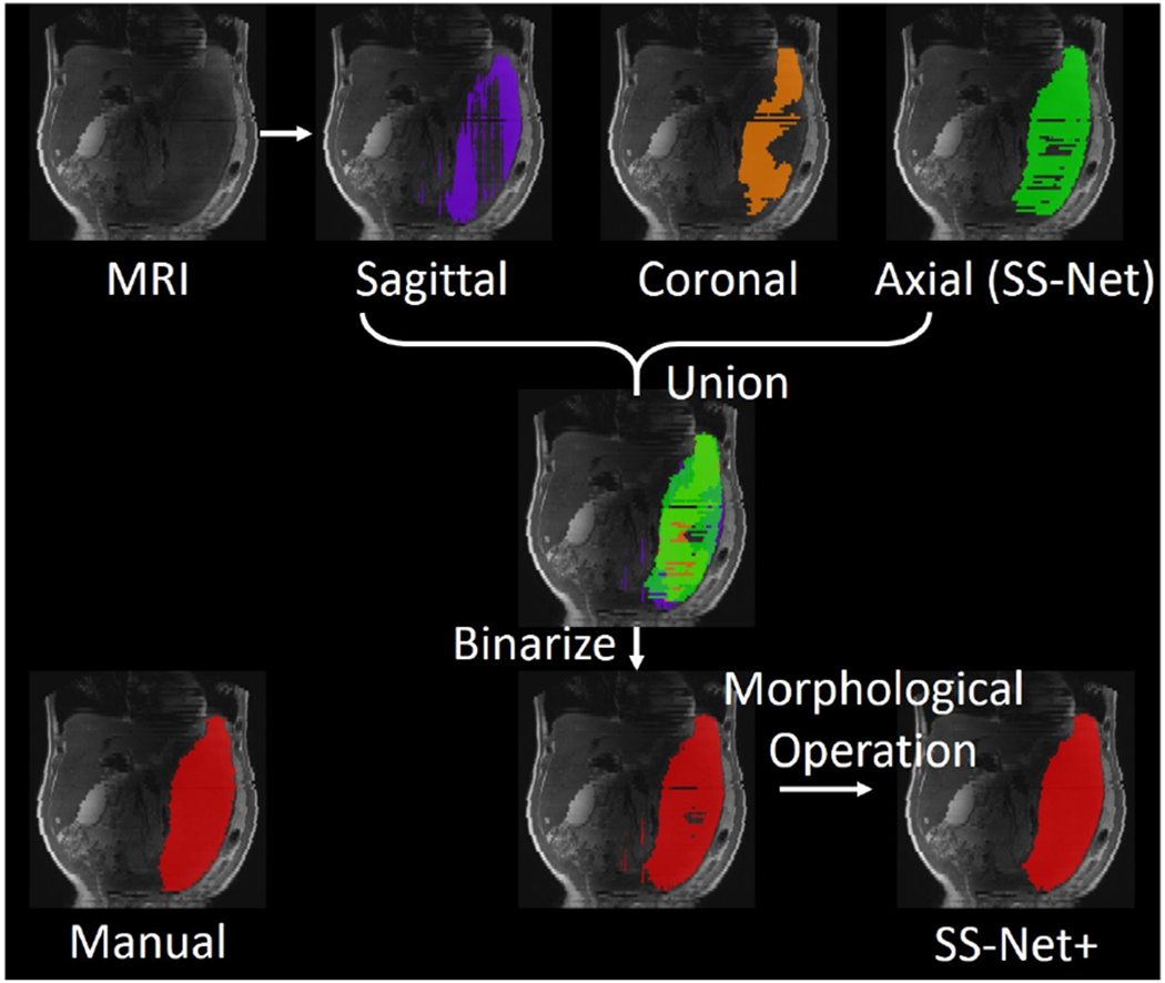
Fig. 6.

This figure presented the experimental design of the multi-view fusion stage (“+” sign in Fig. 5). Three whole spleen segmentations were obtained from input MRI volume. Then, the union operation was used to combine three segmentation to a single whole spleen segmentation. Last, the binary 3D morphological operations (open and close) were performed to refine the final segmentation result.
