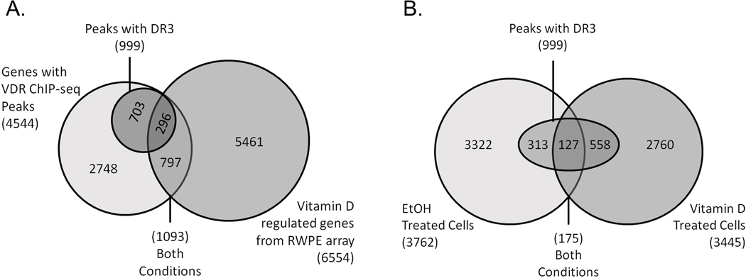
Fig. 5.
Identification of VDR binding sites in RWPE1 cells. (A) A comparison of ethanol vehicle (EtOH) or 1,25(OH)2D (Vitamin D) treated cells (10 nM, 3 h). Traditional DR3-type VDR binding motifs were identified below the summits of VDR peaks. (B) Peaks were assigned to genes and then compared to transcriptome results from vitamin D treated RWPE1 cells. Overlap among the groups and the DR3 containing VDR peaks was assessed.

