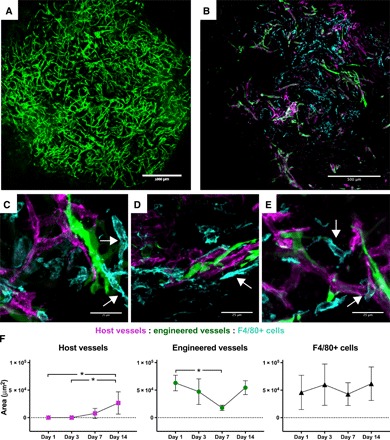
Fig. 4. Macrophage interaction with host and engineered vasculature within the graft.

(A) Representative image of an engineered vascular graft bearing GFP-expressing human blood vessels (green) after 14 days of in vitro culture. Scale bar, 1000 μm. (B) F4/80 immunostaining (cyan) of engineered vascular graft explanted 14 days after implantation, indicating interactions between engineered vessel network (green) and penetrating host vessels (red). Scale bar, 500 μm. (C to E) High-magnification images of macrophages (F4/80+, cyan) interacting with engineered (green) and host (magenta) vessels within graft marked with white arrows. Scale bars, 25 μm. (C) Macrophages form vessel-like structures; (D) macrophage adjacent to both engineered and host vessel; (E) macrophage bridging between vessel segments and wrapping around host vessel. (F) Quantification of the area of host vessels, engineered vessels, and F4/80+ cells in sectioned grafts explanted on days 1, 3, 7, and 14. Data represent means ± SD and were analyzed using one-way ANOVA with Tukey’s post hoc multiple comparisons test; *P < 0.05 for n = 6 to 8.
