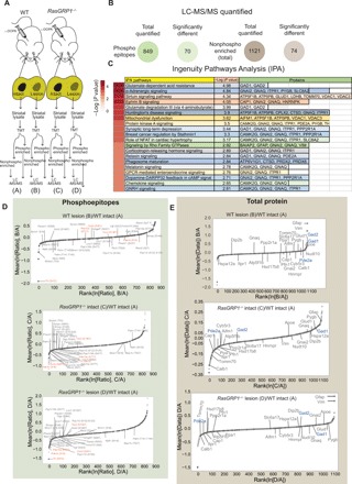
Fig. 5. Quantitative proteomics of the striatum of WT and RasGRP1 KO dyskinesia animals.

(A) Scheme of isolation of striatal tissue from the 6-OHDA–lesioned WT and RasGRP1 KO (RasGRP1−/−) after l-DOPA treatment, followed by LC-MS/MS. (B) Total number of quantifiable proteins that are enriched for phosphorylated epitopes and nonphosphorylated total protein. (C) Ingenuity Pathway Analysis (IPA) analysis for significantly altered nonphosphorylated proteins. Relative quantitation of phosphopeptide (D) and non-phosphopeptide [total protein; (E)] abundance between WT lesion/ WT intact, RasGRP1 KO intact/ WT intact and l RasGRP1 KO lesion/WT intact groups. Significant targets and nonsignificant targets were indicated in dark and light gray circles, respectively (n = 3 mice per group).
