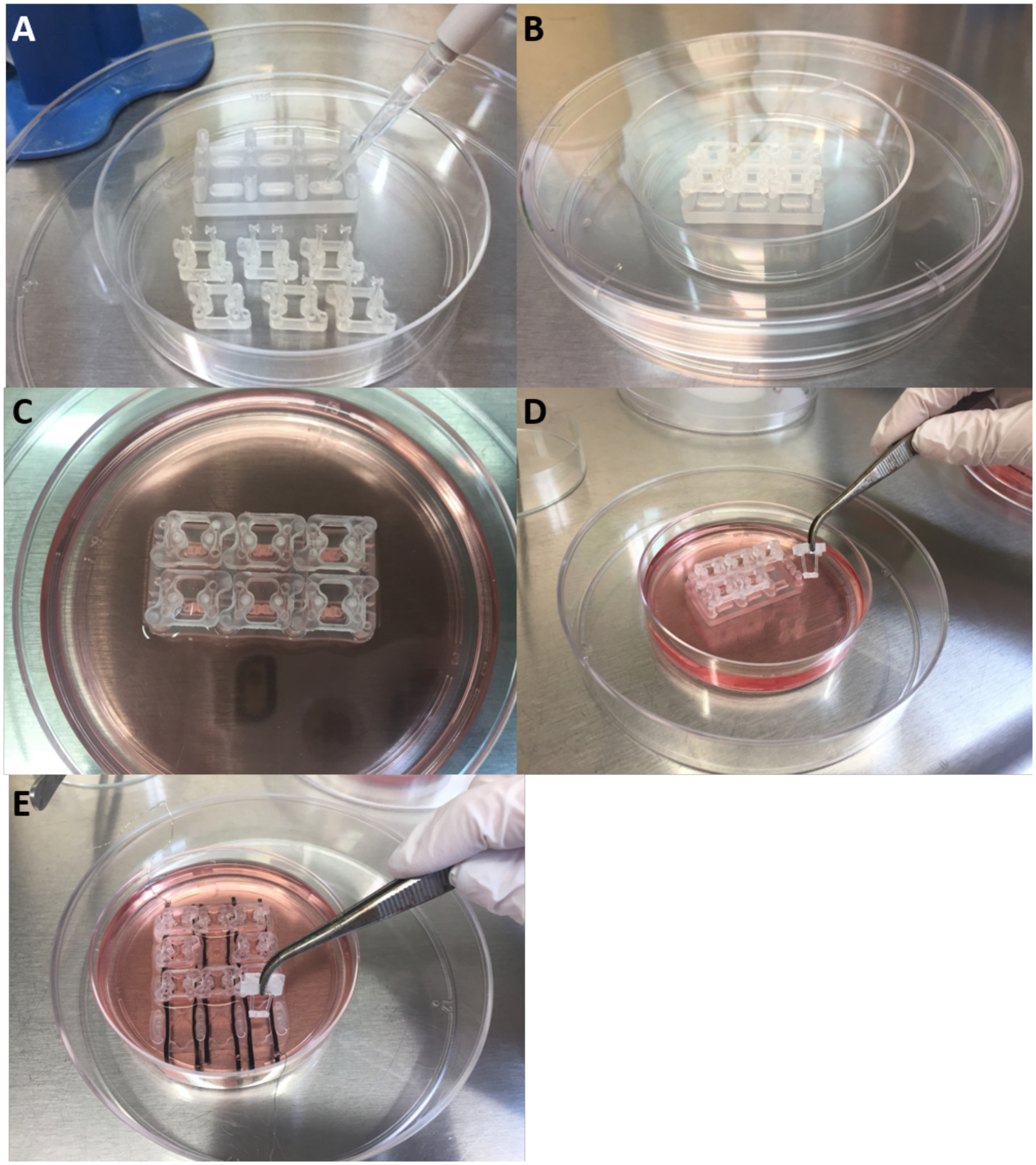
Figure 6. Overview of cardiac tissue formation.

A) Hydrophobic coating of tissue formation chamber platform. B) PDMS pillars are placed back into tissue formation reactor after coating. C) The cardiac tissue is formed around the pillars and let compact over 7 days. D-E) The cardiac tissues are transferred to the cardiac maturation platform.
