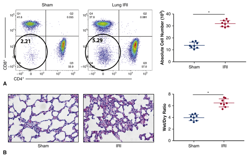
Figure 2.
Rapid Expansion of DN T cells following lung IRI. A left dorsolateral thoracotomy was performed in the fourth intercostal space and a microvascular clamp was applied across the pulmonary artery and vein to occlude the vasculature. The left lung was occluded for 30 minutes, and the microvascular clamp was removed. The thoracotomy was closed and the mouse was removed from the ventilator and allowed to recover for the 3-hour reperfusion period. Mice were sacrificed, underwent midline abdominal incisions, were exsanguinated, and had their lung lobes removed for lymphocyte collection. (A) Mice that underwent lung IRI demonstrated an approximately 2-fold rise in DN T cells in the lungs (5.29% ± 1%, p< 0.01) when compared with surgical sham controls (2.21% ± 3%, p<0.01). (C) Illustrates absolute number of DN T cells and it is a cumulative data on eight independent experiments (n=8). (B) Hematoxylin and eosin staining showed mice that underwent lung IRI exhibited thickened interstitium and neutrophil infiltration when compared to the sham controls. (D) Shows Wet/Dry ratio, with lungs that underwent IRI exhibiting higher W/D ratio.

