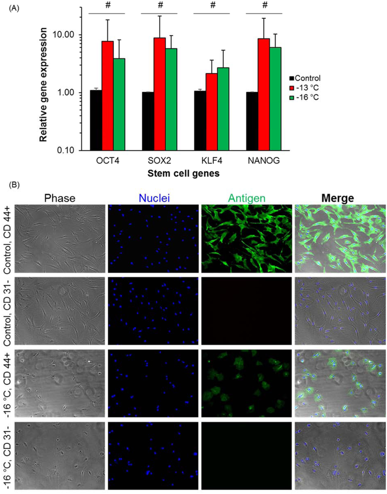
Figure 4.
Stem cell genes expressions and immunofluorescence staining of surface protein markers. (A) Gene expression levels examined by RT-PCR. Four stem cell genes OCT 4, SOX2, KLF4 and NANOG for ADSCs were examined. Number of independent experiments n = 3, #: p > 0.1. (B) Immunofluorescence staining for stem cell surface markers. Both positive (CD 44+) and negative (CD 31 −) surface markers were examined for fresh control and DSC preserved cells at −16 °C for 7 days.

