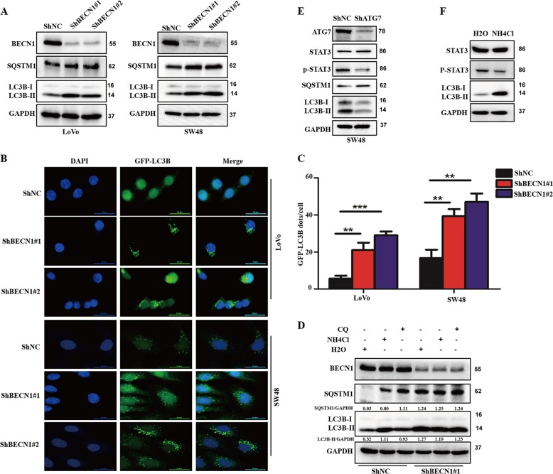
Fig. 5. Knockdown of BECN1 decreases autophagic flux in colorectal cancer.
a Western blot analysis used to determine the protein levels of LC3B, BECN1, SQSTM1/p62, and GAPDH in both LoVo and SW48 cells expressing the negative control RNA or shRNA-BECN1. b, c LoVo and SW48 cells expressing the negative control RNA or shRNA-BECN1 were transfected with GFP-LC3B plasmids for 48 h, and then the GFP-LC3B signal was examined under confocal microscopy. The amount of GFP-LC3B signal in each cell was quantified. Scale bar: 50 μm. d SW48 cells expressing the negative control RNA or shRNA-BECN1#1 were treated with or without 25 μM CQ for 2 h or 5 mM NH4Cl for 4 h. The accumulation of LC3B and SQSTM1was determined by a western blot assay and represented the autophagic flux. The LC3B accumulation and SQSTM1 was quantified using GAPDH as the loading control. The results indicate the mean ± SD for triplicate independent experiments (*P < 0.05, **P < 0.01, ***P < 0.001 by an independent Student’s t test). e SW48 cells were transfected with negative control siRNA (siNC) or siRNA-ATG7 (siATG7) for 72 h, and then cells were harvested for western blot assays. f SW48 cells were treated with H2O or 5 mM NH4Cl for 4 h, and then cells were harvested for western blot assays.

