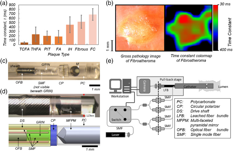
Fig. 3.
(a) Mean computed for different plaque groups under static conditions. The error bars indicate standard error of the mean. TCFA, thick-cap FA (THFA), pathological intimal thickening (PIT), nonnecrotic fibroatheroma (FA), intimal hyperplasia (IH), fibrous plaque, and fibrocalcific plaque (FC). (b) Color-map of the distribution (30 to 400 ms) over ROI across lesion. Presence of lipid-rich plaque with a well-defined outline is clear from the color-map and is backed-up by the corresponding gross pathology photograph (adapted with modifications from Ref. 30). (c) The first-generation LSR endoscope. Reflection of the OFB is seen in the circular flat mirror, and the single-mode illumination (SMF) fiber runs parallel underneath the optical assembly (not visible). Scale bars: 1 mm (adapted with modifications from Ref. 33). (d) The omnidirectional LSR catheter assembly. Distal optics design is optimized for a lumen of 3 mm diameter (size of a human coronary artery). Top: The photograph of the distal optic assembly. The catheter diameter is 1.2 mm. Bottom: The computer-aided drawing shows the catheter distal optics assembly which incorporates SMFs for illumination, a circular polarizer (CP), an MFPM, a gradient-index (GRIN) lens, and an OFB. The distal assembly is housed within a protective polycarbonate (PC) tube and a customized drive shaft (DS). (e) Schematic diagram of the omnidirectional LSR catheter, pull-back assembly, and console hardware. Laser light (633 nm, 22.5 mW) is coupled into an SMF passes through an MEMS switch and is split to four illumination fibers. The speckle patterns obtained from the lumen wall are transmitted through an OFB and are imaged on the CMOS camera [(d) and (e) are adapted with modifications from Ref. 45].

