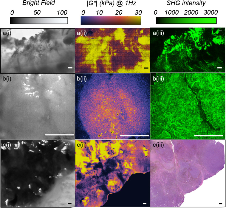
Fig. 6.
Bright-field images, LSR maps, and either of histology or SHG images of human tissue specimens associated with three different cancers, namely: a(i–iii) breast cancer (invasive ductal carcinoma), b(i–iii) squamous cell lung cancer, and c(i–iii) melanoma skin cancer. Strong qualitative agreement is observed between LSR maps of the viscoelastic modulus and the intuitive perception of stiffness from the histopathological and compositional images in all three cases. Regions of increased viscoelastic modulus, in a–c(ii), coincide with fibrous stroma in H&E section and the foci of collagen accumulation and alignment in the SHG images, in a–c(iii). On the other hand, soft regions of low viscoelastic modulus correspond to cell-dense regions in the H&E section and areas depleted of collagen in the SHG image. The LSR microscopic platform provides the unique capability to map the mechanically heterogeneous tumor microenvironment. Scale bars are .

