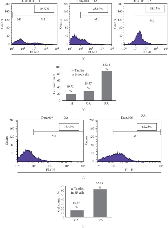
Figure 4.

Validation by FACS. Blood cells or SF cells from RA, OA, and healthy blood cells were isolated and proceeded for FACS analysis with α-Taxilin primary antibody followed by Alexa Fluor 467-tagged secondary antibody. (a) Data captured using FACS caliber showing percentage of cells positively stained with Alexa Fluor 467 in H, OA, and RA blood cells. (b) Graphical representation of FACS data in terms of cells stained with Alexa Fluor for H, OA, and RA samples. (c) Data captured using FACS caliber showing the percentage of cells positively stained with Alexa Fluor 467 in SF cells. (d) Graphical representation of FACS data in terms of cells stained with Alexa Fluor for OA and RA SF cells.
