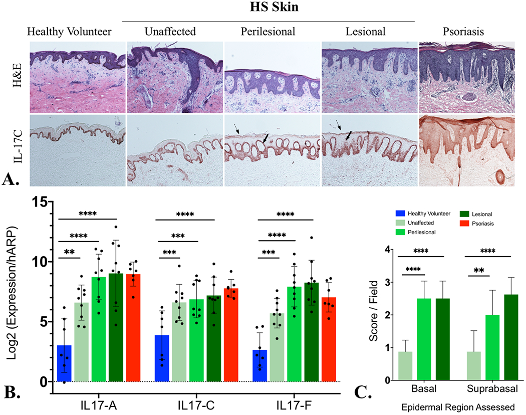
Figure 1:

A) Interleukin 17C (IL-17C) localizes to the keratinocytes of psoriasiform epidermal hyperplasia in Hidradenitis Suppurativa by immunohistochemistry (Fig 1A) with increased IL-17C in suprabasal and granular layer of HS lesional skin. Diffuse dermal and epidermal staining is indicative of IL-17C protein diffusing into the surrounding dermis and epidermis at levels comparable with psoriasis. B) mRNA levels of IL-17C are significantly elevated (using one-way ANOVA) in lesional, perilesional and unaffected skin compared with healthy controls (Fig 1B) and comparable to the levels seen in psoriasis skin (Fig 1B). mRNA elevations of IL-17A and IL-17F are provided (Fig 1B) for comparison. Semiquantitative scoring of IL-17C IHC staining identifies statistically significant elevation (by one-way ANOVA) in perilesional and lesional tissue compared to unaffected tissue (Fig 1C).
Key: *= p>0.05; **= p<0.01; ***=p<0.001; ****= p<0.0001
NB: All statistical tests using one-way ANOVA were adjusted for multiple comparisons using the Benjamini-Hochberg method.
