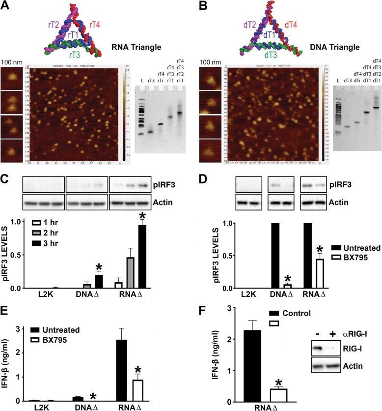
Fig. 5.
RNA triangle nanoparticles stimulate RIG-I dependent responses in hμglia human microglial cells. DNA and RNA triangle formations assessed by atomic force microscopy (AFM) and ethidium bromide total staining native-PAGE (a, b). “L” corresponds to DNA low molecular weight ladder. Cells were untreated or treated with 1 μM BX795, an inhibitor for TBK1/IKKε, for 3 h prior to transfection with 0.1 μg/ml BDNA, 1 μg/ml 5′pppRNA, 5 nM DNA triangles, or 5 nM RNA triangles. Cell lysates collected at 1, 2, or 3 h were analyzed for protein expression of phosphorylated IRF3 (pIRF3) and the housekeeping gene β-actin via immunoblot analysis (c, d). Cell supernatants were collected 24 h post transfection and IFN-β levels were quantified by specific capture ELISA (e). Microglia were treated with scrambled control siRNA or siRNA targeted against RIG-I (αRIG-I) at a final concentration of 5 nM for 24 h. Cells were placed in fresh media for 24 h prior to transfection with 5 nM RNA triangles. Cell supernatants were collected 24 h post transfection and IFN-β levels were quantified by specific capture ELISA. Cell lysates were collected 24 h post transfection and analyzed for protein expression of RIG-I and the housekeeping gene, β-actin, via immunoblot analysis (f). Data are expressed as mean ± SEM for a minimum of three independent experiments. Asterisks indicate statistical significance compared to untreated condition as determined by Student’s t test or two-way ANOVA (p < 0.05)

