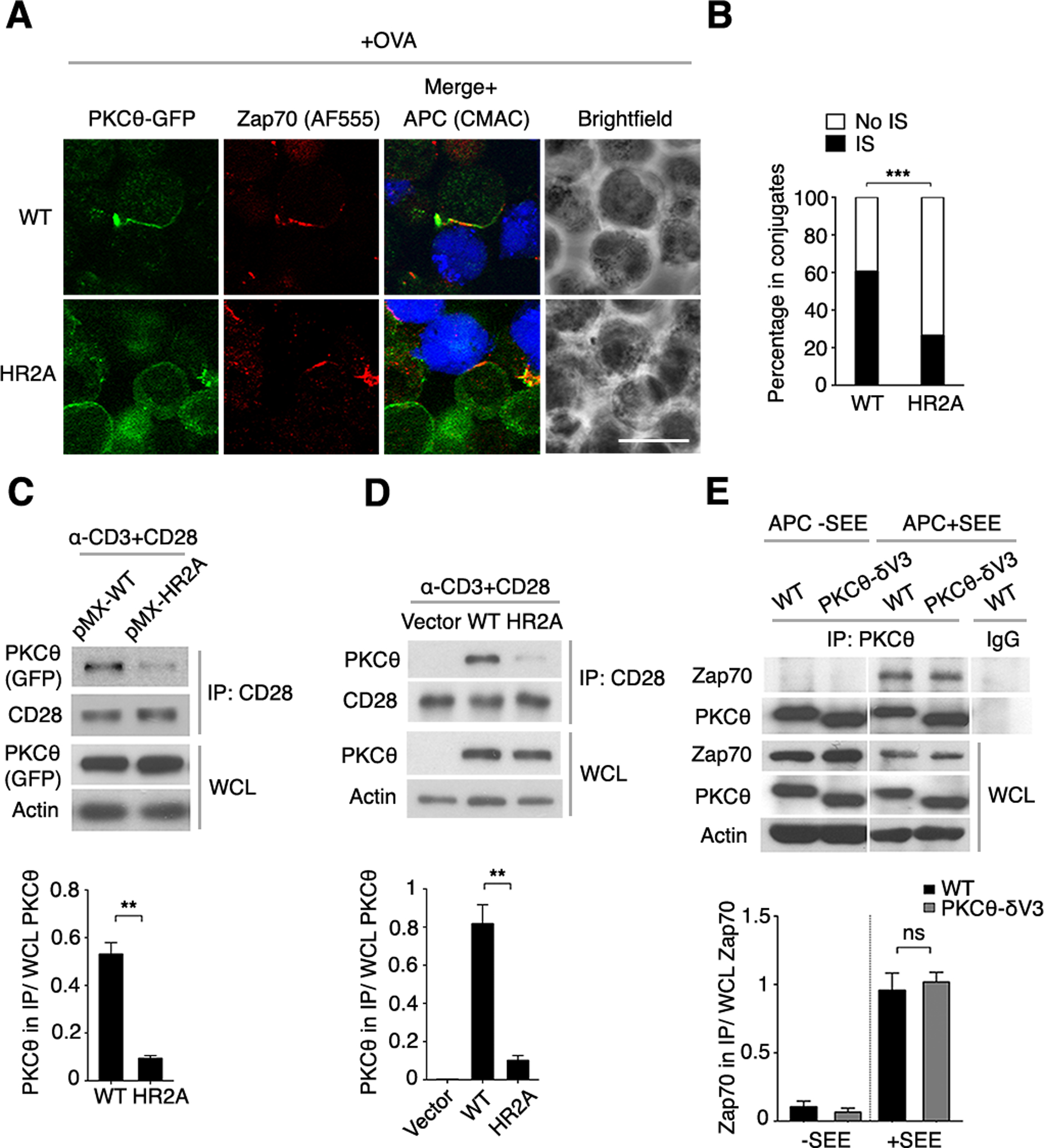
Fig. 6. pTyr binding by PKCθ is required for PKCθ IS localization and association with CD28.

(A) Confocal imaging of Prkcq−/− OT-II CD4+ T cells that were transduced with an RV encoding GFP-tagged WT or HR2A mutant PKCθ (green), mixed with APCs labeled with the cell-tracking dye CMAC (blue), and pulsed with OVA peptide (+OVA). Fixed conjugates were stained for Zap70 plus a secondary fluorescent antibody (AF555). A representative cell is shown. Scale bar, 10 μm.
(B) Quantification of PKCθ localization within (IS) or outside of (No IS) the IS in the T cell-APC conjugates analyzed in (A). Analysis was performed only on conjugates that displayed IS-localized Zap70 and detectable PKCθ. n = ~120.
(C to E) GFP+ CD4+ T cells transduced in vitro (C and D) or derived from BM chimeras reconstituted in vivo (E) with pMIG vectors expressing PKCθWT, PKCθHR2A, or PKCθ-δV3 were stimulated with mAbs against CD3 and CD28 for 5 min. CD28 and PKCθ interaction was detected by co-immunoprecipitation (co-IP). Representative blots are shown above the corresponding band densitometry quantifications pooled from 3 independent experiments. ns, not significant, **P < 0.01, ***P < 0.001.
