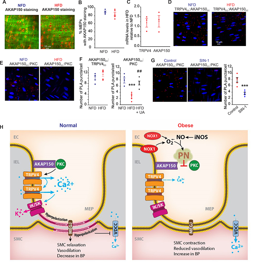
Figure 6. Peroxynitrite (PN) impairs AKAP150EC anchoring of PKC in obesity.
(A) Representative merged images from en face preparations of third-order MAs showing IEL autofluorescence (green) and AKAP150EC immunofluorescence (red) in NFD (left) and HFD mice (right). (B) Quantification of AKAP150EC staining at MEPs in NFD and HFD mice (n = 5). (C) Quantification of TRPV4 and AKAP150 mRNA levels in homogenates of third-order MAs from HFD mice, expressed relative to those in NFD mice (n = 3). (D) Representative PLA merged images of EC nuclei (blue) and AKAP150EC:TRPV4EC co-localization (red puncta) in en face preparations of third-order MAs from NFD (left) and HFD (right) mice. (E) Representative PLA merged images of EC nuclei (blue) and AKAP150EC:PKC co-localization (red puncta) in third-order en face preparations of MAs from NFD (left) and HFD (right) mice. (F) Quantification of AKAP150EC:TRPV4EC co-localization (left) and AKAP150EC:PKC co-localization (right) in NFD and HFD mice. AKAP150EC:PKC co-localization was rescued by uric acid (UA, 200 μM) in HFD mice (n = 5; ***P < 0.001 for HFD only vs. NFD only, #P < 0.01 for UA-treated HFD vs. HFD only; one-way ANOVA). (G) Left, representative PLA merged images of EC nuclei (blue) and AKAP150EC:PKC co-localization (red) in MAs from WT mice in the absence or presence of 50 μM SIN-1. Right, quantification of AKAP150EC:PKC co-localization (right) in WT mice in the absence or presence of SIN-1 (n = 5; P < 0.001 vs. Control; t-test). (H) Schematic depicting the PN-dependent signaling mechanism that impairs endothelial function and elevates blood pressure in obesity.

