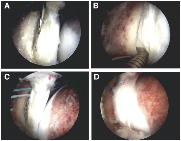
Fig. 4.

Right hip arthroscopy showing the sequence of the repair technique initiating with (A) elevation of the cartilage defect, (B) microfracture beneath through the substrate bone and (C and D) repaired using the adjacent labrum with bone anchors.
