Figure 2.

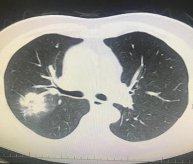
A 66-year-old man’s chest CT imaging, PCR positive, and CT has right upper lobe consolidation which is atypical appearance for COVID-19 pneumonia (archives of Şule Akçay).

A 66-year-old man’s chest CT imaging, PCR positive, and CT has right upper lobe consolidation which is atypical appearance for COVID-19 pneumonia (archives of Şule Akçay).