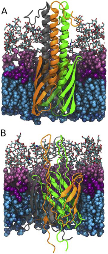
Figure 1:
Comparison between the original YadA-M (PDB: 2LME, [45]) structure (A) and the generated hairpin intermediate (B). The hairpin structure is constructed by stretching the linker and the passenger domain. Lipopolysaccharides in the outer leaflet are shown in pink (with sugar groups in licorice, polar headgroups in light pink and lipid tails in magenta). Phospholipids in the inner leaflet are shown in blue (with lipid tails in cyan and polar heads in dark blue). YadA β-strands are semi-transparent to clearly show the α-helical domain in panel A and the hairpin folding intermediate in panel B. The individual YadA monomers are shown in green, orange, and grey.

