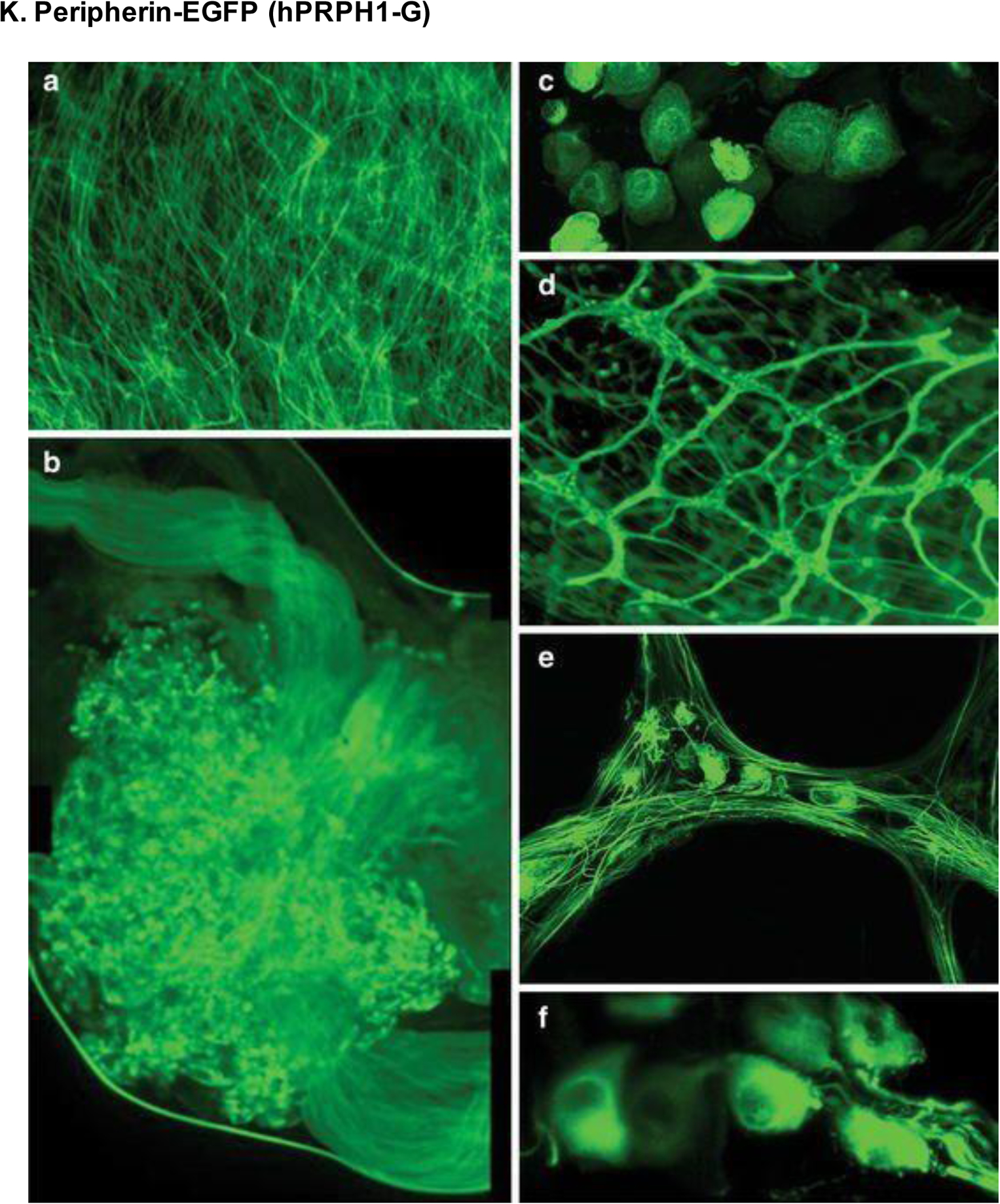
Figure 20.

Expression of peripherin-EGFP (hPRPH1-G) in unfixed tissues viewed by multiphoton and confocal microscopy. (A) Cervical spinal cord at 200× magnification. (B) Images combined to show the DRG. (C) DRG sensory neuron cell bodies. (D-E) Small intestine seen by fluorescence (D) and confocal (E) microscopy at 100× and 400× magnification, respectively. (F) Retinal images showing RGC bodies at 1000× magnification. Reproduced with permission from [220].
