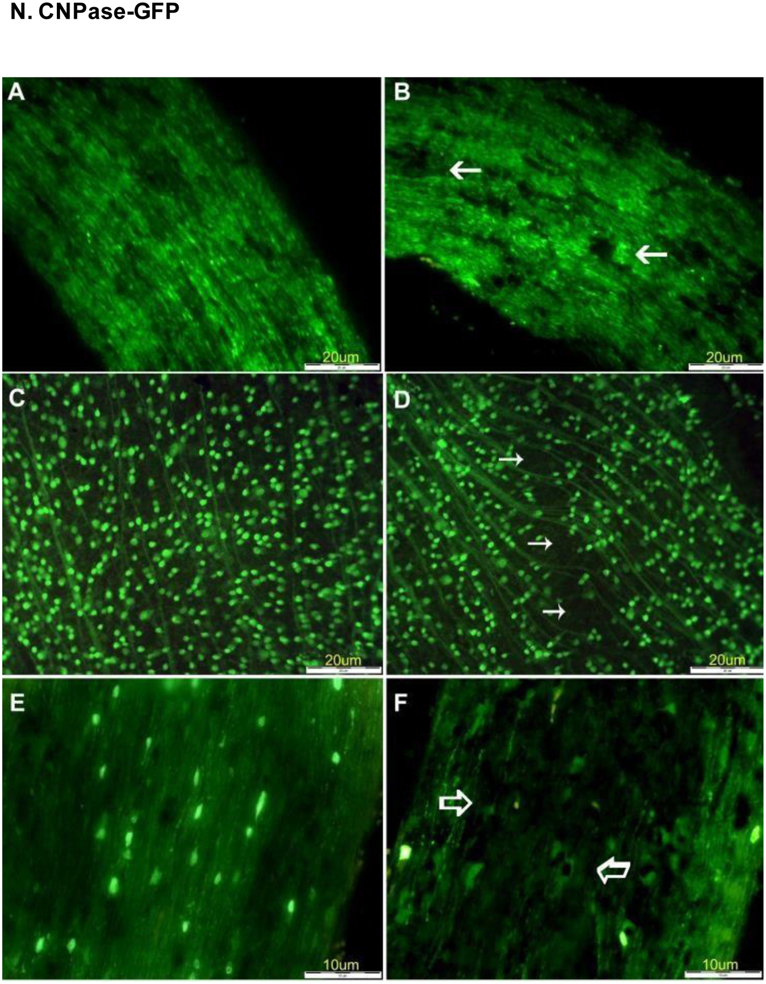
Figure 23.

Expression of Thy1-CFP and CNPase-GFP in the retina and optic nerve of a mouse model of experimentally-induced rodent anterior ischemic optic neuropathy (rAION). (A) Optic nerve images in control Thy1-CFP mice before induction of rAION. (B) View of optic nerve 21 days after induction of rAION, with arrows noting regions of significant axon loss. (C) Retina of control mouse before induction of rAION. (D) Optic nerve of CNPase-GFP mouse before induction of rAION. (F) Loss of oligodendrocyte expression 21 days after induction of rAION. Reproduced with permission from [105].
