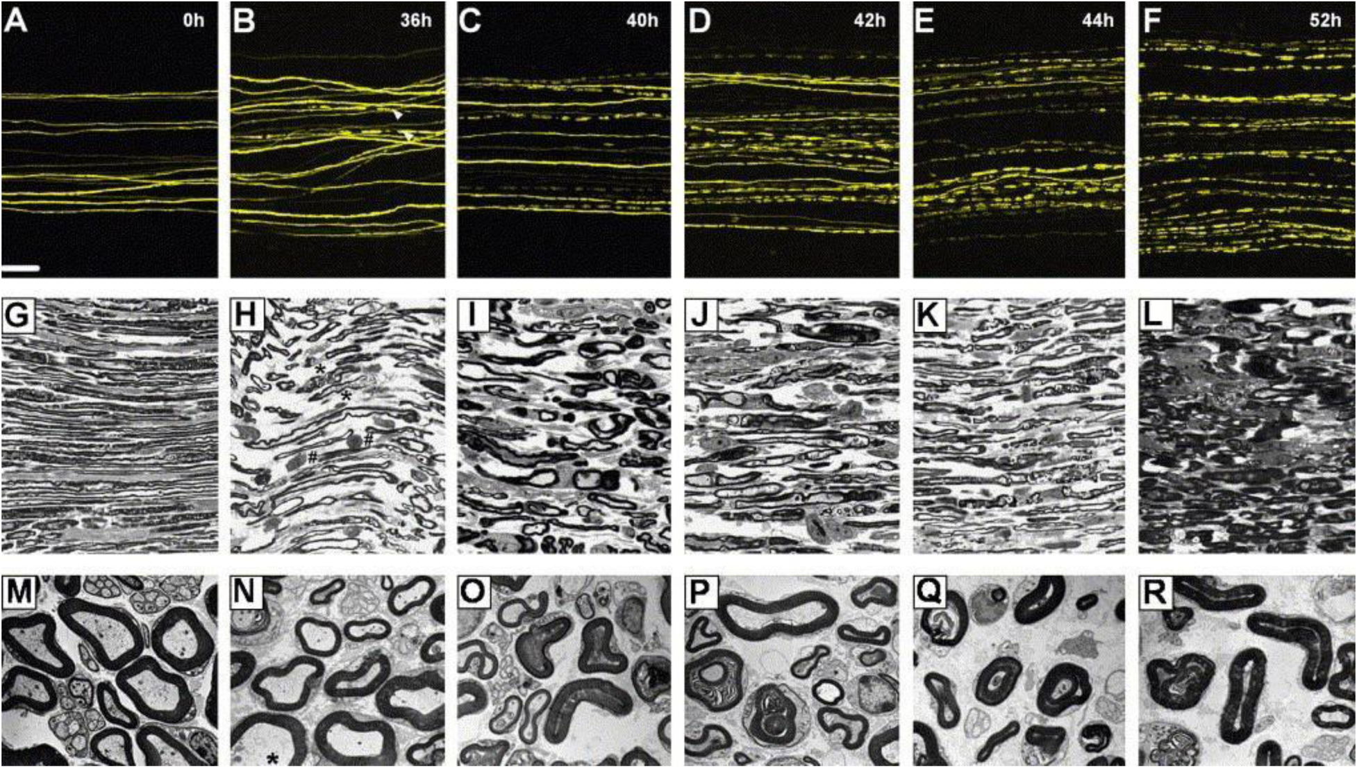
Figure 3.

Imaging of Thy1-YFP+ neurons revealed that YFP axons could be successfully used to display nerve changes following Wallerian degeneration. Such imaging was achieved through light and electron microscopy techniques. Scale bars, (A–F) 100 μm, magnification: (G–L) 1000×, magnification: (M–R) 4400×. Reproduced with permission from [42].
