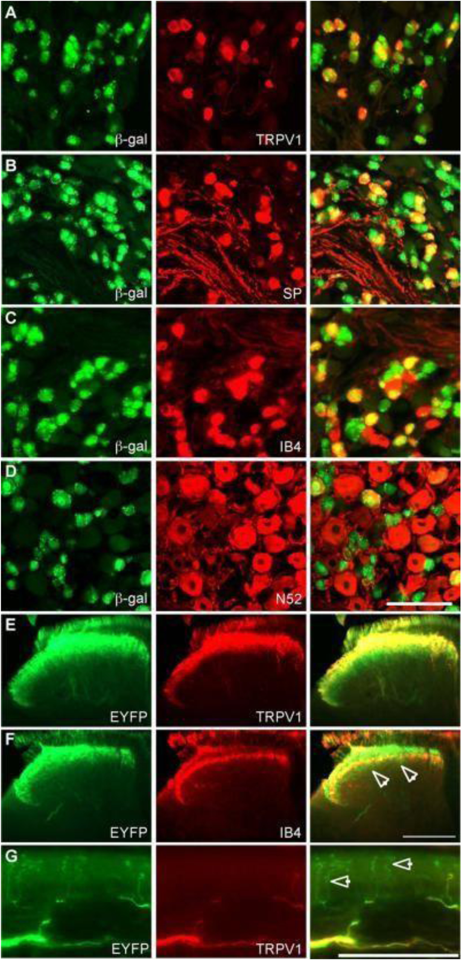
Figure 9.

Staining of DRG and spinal cord from TRPV1Cre mice crossed with multiple Cre-dependent reporter lines. Immunohistochemical staining revealed TRPV1 expression in peptidergic and nonpeptidergic C-fibers, as well as myelinated DRG neurons during development. Scale bars, (D) and (G) 100 μm, (F) 200 μm. Reproduced with permission from [66].
