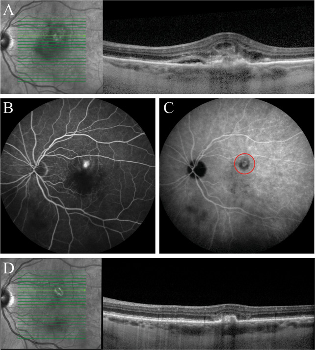
Fig. 2.
Multimodal imaging of an eye of a 73-year-old man diagnosed with polypoidal choroidal vasculopathy who was treated with half-dose photodynamic therapy (PDT) and intravitreal anti-vascular endothelial growth factor (VEGF) injections. a, b Prior to initiation of half-dose PDT, a hyperreflective irregular retinal pigment epithelium layer with adjacent subretinal fluid highly suggestive of a polypoidal lesion was seen on optical coherence tomography (OCT) (a), and leakage of fluorescein was observed on fluorescein angiography (b). The patient had received 2 intravitreal anti-VEGF injections before half-dose PDT. c The mild hyperfluorescent polypoidal lesion visible on indocyanine green angiography was targeted with half-dose PDT (within the red circle). d Compared to the imaging results before PDT, at the visit 6 months after half-dose PDT, the polypoidal lesion had regressed and the subretinal fluid had disappeared on OCT. This patient had received an additional 7 intravitreal anti-VEGF injections between half-dose PDT and the visit at 6 months after PDT

