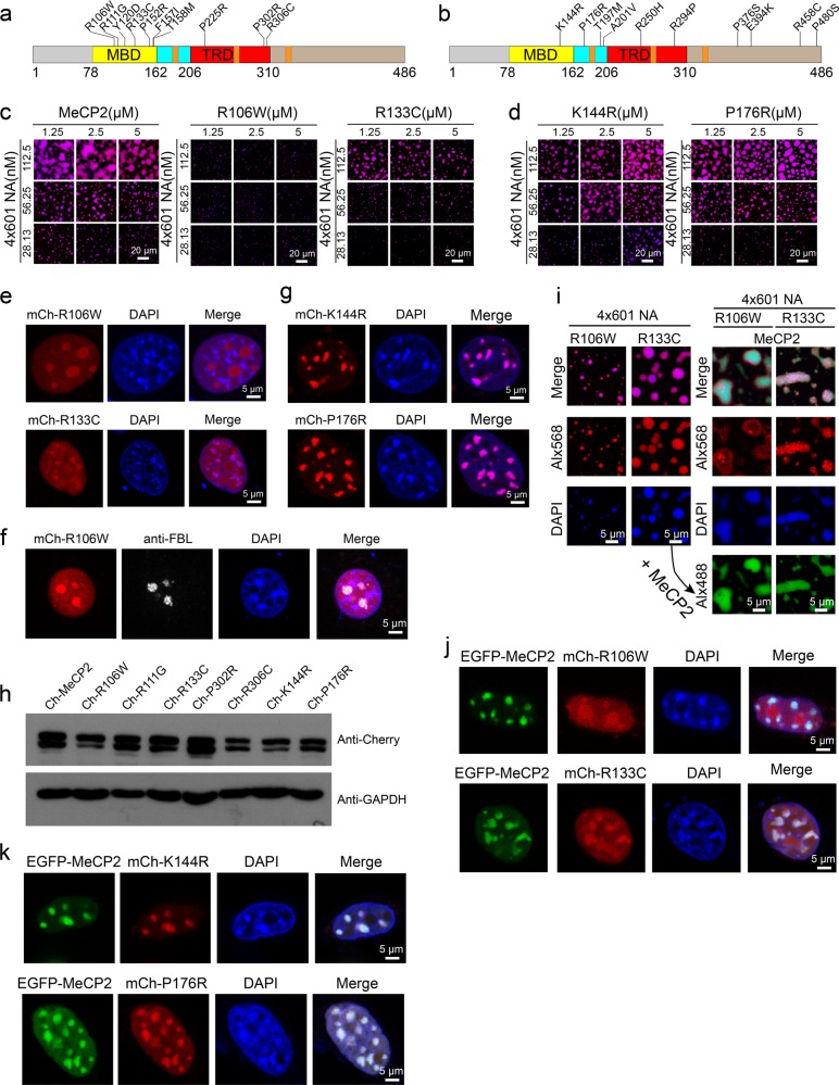
Fig. 4. RTT-related missense mutations in the MBD compromise MeCP2-driven chromatin LLPS.
a, b Schematic diagrams of the missense mutations analyzed in this study. Ten Rett-related MeCP2 missense mutations in MBD or TRD (a). Ten neutral MeCP2 missense variants, which are not associated with disease symptoms (b). c Phase diagrams of WT MeCP2 and two RTT-related missense mutations in MBD (R106W and R133C) with 4×601-NA. Scale bars, 20 µm. d Phase diagrams of MeCP2 benign variants (K144R and P176R) with 4×601-NA. Scale bars, 20 µm. e Dispersion of overexpressed mCherry-MeCP2-R106W or R133C within the nucleus in NIH 3T3 cells. Scale bars, 5 µm. f In NIH 3T3 cells, overexpressed mCherry-MeCP2-R106W is dispersed in the nucleus and also enriched in the nucleolus (labeled with Fibrillarin (FBL)). Scale bar, 5 µm. g Location of overexpressed mCherry-MeCP2-K144R and P176R to heterochromatic chromocenters marked by DAPI in NIH 3T3 cells. Scale bars, 5 µm. h Western blot analysis to check the protein levels of overexpressed WT MeCP2 and MeCP2 mutants in 3T3 cells. Upper panel, anti-Cherry antibody is used. Lower panel, anti-GAPDH antibody is used. GAPDH is used as the internal control. i Left panels, mCherry-MeCP2-R106W and R133C undergo weak phase separation with 4×601-NA. Right panels, phase separation after addition of WT Alx488-MeCP2 into the reactions shown in the left panels. Scale bars, 5 µm. j Co-expression of EGFP-MeCP2 with mCherry-MeCP2-R106W or R133C in NIH 3T3 cells. Scale bars, 5 µm. k Colocalization of EGFP-MeCP2 with mCherry-MeCP2-K144R or P176R in NIH 3T3 cells. Scale bars, 5 µm.

