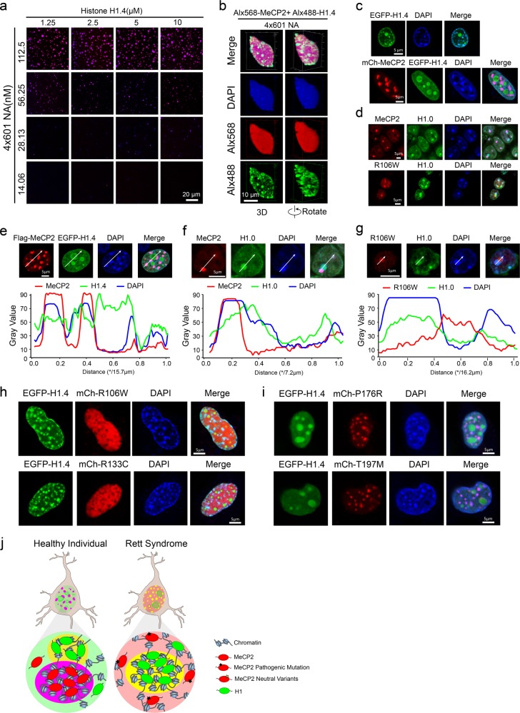
Fig. 6. Competition and incompatibility between MeCP2- and H1-containing chromatin puncta.
a Phase diagram of human linker histone H1.4 with 4×601-NA. Scale bar, 20 µm. b 3D shapes of H1.4-NA droplets within a big MeCP2-NA droplet at a high concentration of 4×601-NA. Scale bar, 10 µm. c Top panel, overexpressed EGFP-H1.4 forms chromatin puncta and colocalizes with heterochromatic chromocenters in NIH 3T3 cells. Bottom panel, co-expression of mCherry-MeCP2 with EGFP-H1.4 in NIH 3T3 cells. mCherry-MeCP2 forms chromatin puncta and colocalizes with heterochromatic chromocenters, whereas H1.4 is dispersed within the nucleus and enriched in the nucleolus. Scale bars, 5 µm. d Top, Immunofluorescence analysis of MeCP2 WT and H1.0 in hippocampal neurons from mice. Bottom, Immunofluorescence analysis of MeCP2 R106W and H1.0 in hippocampal neurons from mice. Scale bars, 5 µm. e Top panel, immunofluorescence images of overexpressed Flag-MeCP2 and EGFP-H1.4 in NIH 3T3 cells, Scale bar, 5 µm. Bottom panel, plots of the red, green and blue pixel intensities along the white arrow in the top panel. f Top panel, immunofluorescence images of MeCP2 and H1.0 in mouse hippocampal neurons. Scale bar, 5 µm. Bottom panel, plots of the red, green and blue pixel intensities along the white arrow in the top panel. g Top panel, immunofluorescence images of MeCP2 R106W mutant and H1.0 in mouse hippocampal neurons. Scale bar, 5 µm. Bottom panel, plots of the red, green and blue pixel intensities along the white arrow in the top panel. h Overexpression of EGFP-H1.4 with the MeCP2 mutants, mCherry-MeCP2 R106W or R133C in NIH 3T3 cells. MeCP2 mutant proteins are dispersed within the nucleus in cells, whereas H1 localizes to heterochromatin. Scale bars, 5 µm. i Overexpression of EGFP-H1.4 with two polymorphic MeCP2 variants, mCherry-MeCP2-P176R or T197M, in NIH 3T3 cells. The MeCP2 benign variants localize to heterochromatin, while H1 mainly localizes within the nucleolus. Scale bars, 5 µm. j Model of the phase distribution of H1 and MeCP2 in neurons. Left, H1 and MeCP2 phases are mutually exclusive but close to each other in healthy neurons. Right, MeCP2 proteins harboring MBD RTT mutations are dispersed in the nucleus, while H1 still forms phase-separated compartments in diseased neurons.

