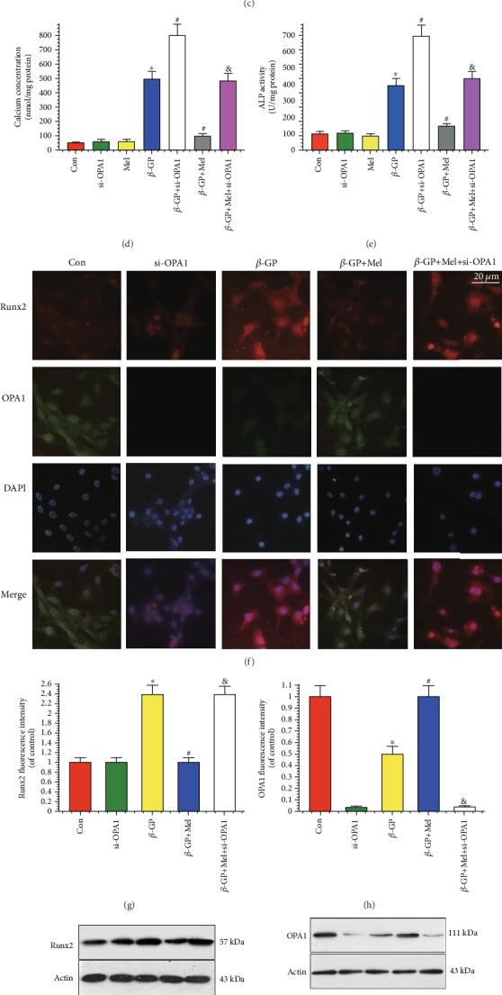
Figure 1.

Melatonin reduced β-GP-induced calcium deposition via OPA1 in VSMCs (n = 6/group). VSMCs were cultured with Dulbecco's Modified Eagle's Medium containing 10% fetal bovine serum and 10 mM β-GP for 14 days. (a, b) Results of different concentrations of melatonin on calcium content and alkaline phosphatase (ALP) activity. (c) Result of melatonin (5 μM) on Alizarin Red S staining. (d) Result of calcium concentration. (e) Result of ALP activity. (f–h) Result of immunofluorescence assay (red signal represents Runx2, green signal represents OPA1). (i, j) Results of Runx2 and OPA1 protein expression. ∗P < 0.05 vs. Con, #P < 0.05 vs. β-GP, and &P < 0.05 vs. β-GP+Mel.
