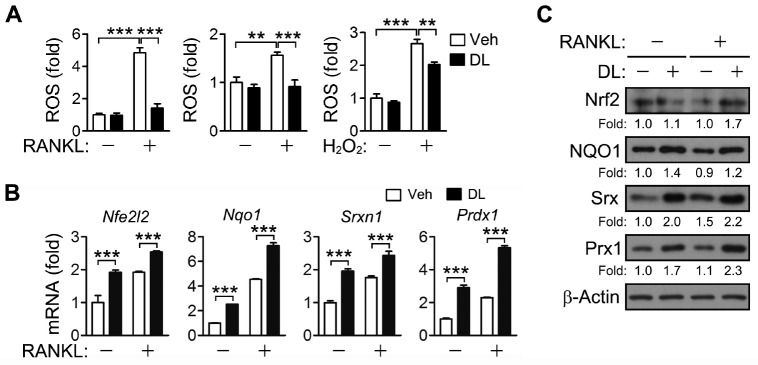
Fig. 3.
ROS removal and Nrf2 activation by DL. (A) BMMs were incubated with RANKL for 2 days (left) and 15 min (center), or with 100 μM H2O2 for 15 min (right) in the presence of 1.5 μM DL. The cells were incubated with 5 μM 5-(and-6-)chloromethyl-2’,7’-dichlorodihydrofluorescein diacetate. The cells were analyzed by a flow cytometer, and the relative levels of fluorescence were presented as fold differences. (B, C) BMMs were treated with RANKL for 24 h in the presence of 1.5 μM DL. The mRNA and protein levels of Nrf2 and its target genes were assessed by real-time PCR (B) and immunoblotting (C), respectively. All values represent means ± SD. N = 3. **P < 0.01; ***P < 0.001 between the indicated groups.

